3R0W

Crystal Structures of Multidrug-resistant HIV-1 Protease in Complex with Mechanism-Based Aspartyl Protease Inhibitors.


Experimental Data Snapshot

  • Method: X-RAY DIFFRACTION
  • Resolution: 1.70 Å
  • R-Value Free: 0.245 
  • R-Value Work: 0.198 
  • R-Value Observed: 0.200 

wwPDB Validation   3D Report Full Report


Ligand Structure Quality Assessment 


This is version 1.4 of the entry. See complete history


Literature

Crystal structures of multidrug-resistant HIV-1 protease in complex with two potent anti-malarial compounds.

Yedidi, R.S.Liu, Z.Wang, Y.Brunzelle, J.S.Kovari, I.A.Woster, P.M.Kovari, L.C.Gupta, D.

(2012) Biochem Biophys Res Commun 421: 413-417

  • DOI: https://doi.org/10.1016/j.bbrc.2012.03.096
  • Primary Citation of Related Structures:  
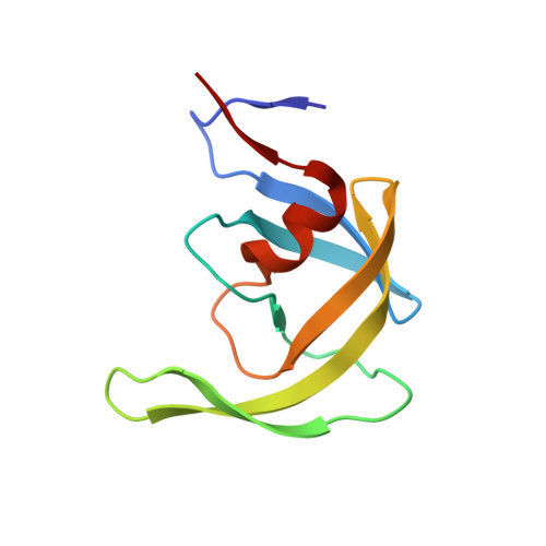
    3R0W, 3R0Y

  • PubMed Abstract: 

    Two potent inhibitors (compounds 1 and 2) of malarial aspartyl protease, plasmepsin-II, were evaluated against wild type (NL4-3) and multidrug-resistant clinical isolate 769 (MDR) variants of human immunodeficiency virus type-1 (HIV-1) aspartyl protease. Enzyme inhibition assays showed that both 1 and 2 have better potency against NL4-3 than against MDR protease. Crystal structures of MDR protease in complex with 1 and 2 were solved and analyzed. Crystallographic analysis revealed that the MDR protease exhibits a typical wide-open conformation of the flaps (Gly48 to Gly52) causing an overall expansion in the active site cavity, which, in turn caused unstable binding of the inhibitors. Due to the expansion of the active site cavity, both compounds showed loss of direct contacts with the MDR protease compared to the docking models of NL4-3. Multiple water molecules showed a rich network of hydrogen bonds contributing to the stability of the ligand binding in the distorted binding pockets of the MDR protease in both crystal structures. Docking analysis of 1 and 2 showed a decrease in the binding affinity for both compounds against MDR supporting our structure-function studies. Thus, compounds 1 and 2 show promising inhibitory activity against HIV-1 protease variants and hence are good candidates for further development to enhance their potency against NL4-3 as well as MDR HIV-1 protease variants.


  • Organizational Affiliation

    Department of Biochemistry and Molecular Biology, School of Medicine, Wayne State University, 540 E. Canfield Avenue, Detroit, MI 48201, USA. yedidirs@mail.nih.gov


Macromolecules
Find similar proteins by:  (by identity cutoff)  |  3D Structure
Entity ID: 1
MoleculeChains Sequence LengthOrganismDetailsImage
Multidrug-resistant clinical isolate 769 HIV-1 Protease
A, B
99Human immunodeficiency virus 1Mutation(s): 0 
UniProt
Find proteins for Q000H7 (Human immunodeficiency virus 1)
Explore Q000H7 
Go to UniProtKB:  Q000H7
Entity Groups  
Sequence Clusters30% Identity50% Identity70% Identity90% Identity95% Identity100% Identity
UniProt GroupQ000H7
Sequence Annotations
Expand
  • Reference Sequence
Small Molecules
Ligands 1 Unique
IDChains Name / Formula / InChI Key2D Diagram3D Interactions
RSY
Query on RSY

Download Ideal Coordinates CCD File 
C [auth A]N-[(2R)-1-{[(2S,3S)-5-{[(2R)-1-{[(2S)-1-amino-4-methyl-1-oxopentan-2-yl]amino}-3-chloro-1-oxopropan-2-yl]amino}-3-hydroxy-5-oxo-1-phenylpentan-2-yl]amino}-3-methyl-1-oxobutan-2-yl]pyridine-2-carboxamide
C31 H43 Cl N6 O6
AWQGEUFVOMSATO-HOMAGXEVSA-N
Binding Affinity Annotations 
IDSourceBinding Affinity
RSY PDBBind:  3R0W IC50: 3.00e+4 (nM) from 1 assay(s)
Binding MOAD:  3R0W IC50: 3.03e+4 (nM) from 1 assay(s)
Experimental Data & Validation

Experimental Data

  • Method: X-RAY DIFFRACTION
  • Resolution: 1.70 Å
  • R-Value Free: 0.245 
  • R-Value Work: 0.198 
  • R-Value Observed: 0.200 
  • Space Group: P 41
Unit Cell:
Length ( Å )Angle ( ˚ )
a = 45.727α = 90
b = 45.727β = 90
c = 102.149γ = 90
Software Package:
Software NamePurpose
HKL-2000data collection
MOLREPphasing
REFMACrefinement
HKL-2000data reduction
SCALEPACKdata scaling

Structure Validation

View Full Validation Report


Ligand Structure Quality Assessment 


Entry History 

Deposition Data

Revision History  (Full details and data files)

  • Version 1.0: 2012-04-04
    Type: Initial release
  • Version 1.1: 2012-04-25
    Changes: Database references
  • Version 1.2: 2012-05-23
    Changes: Database references
  • Version 1.3: 2012-12-12
    Changes: Other
  • Version 1.4: 2023-09-13
    Changes: Data collection, Database references, Derived calculations, Refinement description