3QIS

Recognition of the F&H motif by the Lowe Syndrome protein OCRL


Experimental Data Snapshot

  • Method: X-RAY DIFFRACTION
  • Resolution: 2.30 Å
  • R-Value Free: 0.238 
  • R-Value Work: 0.195 
  • R-Value Observed: 0.198 

wwPDB Validation   3D Report Full Report


This is version 1.4 of the entry. See complete history


Literature

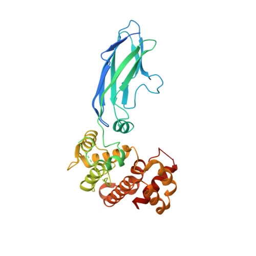
Recognition of the F&H motif by the Lowe syndrome protein OCRL.

Pirruccello, M.Swan, L.E.Folta-Stogniew, E.De Camilli, P.

(2011) Nat Struct Mol Biol 18: 789-795

  • DOI: https://doi.org/10.1038/nsmb.2071
  • Primary Citation of Related Structures:  
    3QIS

  • PubMed Abstract: 

    Lowe syndrome and type 2 Dent disease are caused by defects in the inositol 5-phosphatase OCRL. Most missense mutations in the OCRL ASH-RhoGAP domain that are found in affected individuals abolish interactions with the endocytic adaptors APPL1 and Ses (both Ses1 and Ses2), which bind OCRL through a short phenylalanine and histidine (F&H) motif. Using X-ray crystallography, we have identified the F&H motif binding site on the RhoGAP domain of OCRL. Missense mutations associated with disease affected F&H binding indirectly by destabilizing the RhoGAP fold. By contrast, a disease-associated mutation that does not perturb F&H binding and ASH-RhoGAP stability disrupted the interaction of OCRL with Rab5. The F&H binding site of OCRL is conserved even in species that do not have an identified homolog for APPL or Ses. Our study predicts the existence of other OCRL binding partners and shows that the perturbation of OCRL interactions has a crucial role in disease.


  • Organizational Affiliation

    Department of Cell Biology, Yale University School of Medicine, New Haven, Connecticut, USA.


Macromolecules
Find similar proteins by:  (by identity cutoff)  |  3D Structure
Entity ID: 1
MoleculeChains Sequence LengthOrganismDetailsImage
Inositol polyphosphate 5-phosphatase OCRL-1366Homo sapiensMutation(s): 0 
Gene Names: INPP5FOCRLOCRL1
EC: 3.1.3.36
UniProt & NIH Common Fund Data Resources
Find proteins for Q01968 (Homo sapiens)
Explore Q01968 
Go to UniProtKB:  Q01968
PHAROS:  Q01968
GTEx:  ENSG00000122126 
Entity Groups  
Sequence Clusters30% Identity50% Identity70% Identity90% Identity95% Identity100% Identity
UniProt GroupQ01968
Sequence Annotations
Expand
  • Reference Sequence

Find similar proteins by:  Sequence   |   3D Structure  

Entity ID: 2
MoleculeChains Sequence LengthOrganismDetailsImage
Protein FAM109A13Homo sapiensMutation(s): 0 
UniProt & NIH Common Fund Data Resources
Find proteins for Q8N4B1 (Homo sapiens)
Explore Q8N4B1 
Go to UniProtKB:  Q8N4B1
PHAROS:  Q8N4B1
GTEx:  ENSG00000198324 
Entity Groups  
Sequence Clusters30% Identity50% Identity70% Identity90% Identity95% Identity100% Identity
UniProt GroupQ8N4B1
Sequence Annotations
Expand
  • Reference Sequence
Experimental Data & Validation

Experimental Data

  • Method: X-RAY DIFFRACTION
  • Resolution: 2.30 Å
  • R-Value Free: 0.238 
  • R-Value Work: 0.195 
  • R-Value Observed: 0.198 
  • Space Group: P 31 2 1
Unit Cell:
Length ( Å )Angle ( ˚ )
a = 73.478α = 90
b = 73.478β = 90
c = 145.294γ = 120
Software Package:
Software NamePurpose
DENZOdata reduction
SCALEPACKdata scaling
PHASERphasing
PHENIXrefinement
PDB_EXTRACTdata extraction
CrystalCleardata collection
HKL-2000data reduction
HKL-2000data scaling

Structure Validation

View Full Validation Report


Entry History 

Deposition Data

Revision History  (Full details and data files)

  • Version 1.0: 2011-06-15
    Type: Initial release
  • Version 1.1: 2011-07-13
    Changes: Version format compliance
  • Version 1.2: 2011-07-20
    Changes: Database references
  • Version 1.3: 2017-11-08
    Changes: Advisory, Refinement description
  • Version 1.4: 2023-09-13
    Changes: Advisory, Data collection, Database references, Refinement description