3QI4

Crystal structure of PDE9A(Q453E) in complex with IBMX


Experimental Data Snapshot

  • Method: X-RAY DIFFRACTION
  • Resolution: 2.50 Å
  • R-Value Free: 0.239 
  • R-Value Work: 0.213 
  • R-Value Observed: 0.213 

wwPDB Validation   3D Report Full Report


Ligand Structure Quality Assessment 


This is version 1.2 of the entry. See complete history


Literature

Structural asymmetry of phosphodiesterase-9, potential protonation of a glutamic Acid, and role of the invariant glutamine.

Hou, J.Xu, J.Liu, M.Zhao, R.Luo, H.B.Ke, H.

(2011) PLoS One 6: e18092-e18092

  • DOI: https://doi.org/10.1371/journal.pone.0018092
  • Primary Citation of Related Structures:  
    3QI3, 3QI4

  • PubMed Abstract: 

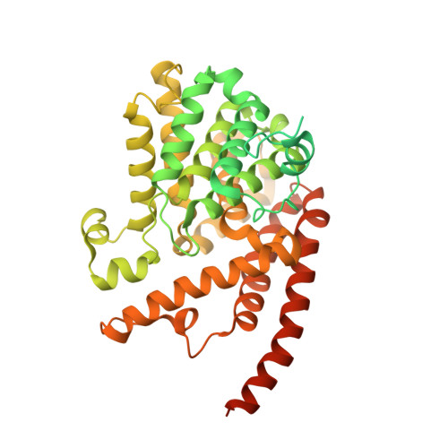
    PDE9 inhibitors show potential for treatment of diseases such as diabetes. To help with discovery of PDE9 inhibitors, we performed mutagenesis, kinetic, crystallographic, and molecular dynamics analyses on the active site residues of Gln453 and its stabilizing partner Glu406. The crystal structures of the PDE9 Q453E mutant (PDE9Q453E) in complex with inhibitors IBMX and (S)-BAY73-6691 showed asymmetric binding of the inhibitors in two subunits of the PDE9Q453E dimer and also the significant positional change of the M-loop at the active site. The kinetic analysis of the Q453E and E406A mutants suggested that the invariant glutamine is critical for binding of substrates and inhibitors, but is unlikely to play a key role in the differentiation between substrates of cGMP and cAMP. The molecular dynamics simulations suggest that residue Glu406 may be protonated and may thus explain the hydrogen bond distance between two side chain oxygens of Glu453 and Glu406 in the crystal structure of the PDE9Q453E mutant. The information from these studies may be useful for design of PDE9 inhibitors.


  • Organizational Affiliation

    Structural Biology Lab, School of Pharmaceutical Sciences, Sun Yat-sen University, Guangzhou, People's Republic of China.


Macromolecules
Find similar proteins by:  (by identity cutoff)  |  3D Structure
Entity ID: 1
MoleculeChains Sequence LengthOrganismDetailsImage
High affinity cGMP-specific 3',5'-cyclic phosphodiesterase 9A
A, B
533Homo sapiensMutation(s): 1 
Gene Names: PDE9A
EC: 3.1.4.35
UniProt & NIH Common Fund Data Resources
Find proteins for O76083 (Homo sapiens)
Explore O76083 
Go to UniProtKB:  O76083
PHAROS:  O76083
GTEx:  ENSG00000160191 
Entity Groups  
Sequence Clusters30% Identity50% Identity70% Identity90% Identity95% Identity100% Identity
UniProt GroupO76083
Sequence Annotations
Expand
  • Reference Sequence
Experimental Data & Validation

Experimental Data

  • Method: X-RAY DIFFRACTION
  • Resolution: 2.50 Å
  • R-Value Free: 0.239 
  • R-Value Work: 0.213 
  • R-Value Observed: 0.213 
  • Space Group: P 41 21 2
Unit Cell:
Length ( Å )Angle ( ˚ )
a = 103.409α = 90
b = 103.409β = 90
c = 269.95γ = 90
Software Package:
Software NamePurpose
AMoREphasing
CNSrefinement
HKL-2000data reduction
SCALEPACKdata scaling

Structure Validation

View Full Validation Report


Ligand Structure Quality Assessment 


Entry History 

Deposition Data

Revision History  (Full details and data files)

  • Version 1.0: 2011-04-27
    Type: Initial release
  • Version 1.1: 2011-07-13
    Changes: Version format compliance
  • Version 1.2: 2024-02-21
    Changes: Data collection, Database references, Derived calculations