


Heme Ruffling Enables the Catalytic Activity of the Heme Degrading Enzyme IsdI
Takayama, S.J., Ukpabi, G.N., Murphy, M.E.P., Mauk, A.G.To be published.
Experimental Data Snapshot
Entity ID: 1 | |||||
|---|---|---|---|---|---|
| Molecule | Chains | Sequence Length | Organism | Details | Image |
| Heme-degrading monooxygenase isdI | 110 | Staphylococcus aureus subsp. aureus N315 | Mutation(s): 0 Gene Names: isdI, SA0160 EC: 1.14.99.3 | 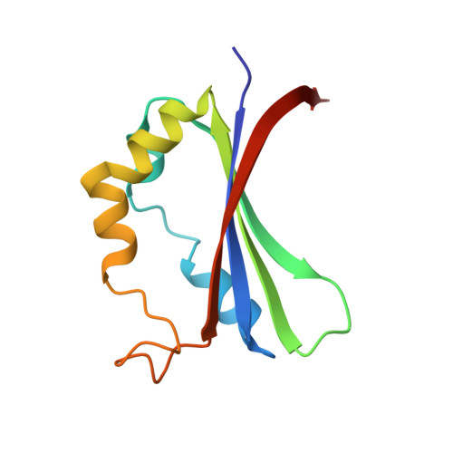 | |
UniProt | |||||
Find proteins for Q7A827 (Staphylococcus aureus (strain N315)) Explore Q7A827 Go to UniProtKB: Q7A827 | |||||
Entity Groups | |||||
| Sequence Clusters | 30% Identity50% Identity70% Identity90% Identity95% Identity100% Identity | ||||
| UniProt Group | Q7A827 | ||||
Sequence AnnotationsExpand | |||||
| |||||
| Ligands 3 Unique | |||||
|---|---|---|---|---|---|
| ID | Chains | Name / Formula / InChI Key | 2D Diagram | 3D Interactions | |
| HEM Query on HEM | C [auth A], E [auth B] | PROTOPORPHYRIN IX CONTAINING FE C34 H32 Fe N4 O4 KABFMIBPWCXCRK-RGGAHWMASA-L |  | ||
| CYN Query on CYN | D [auth A], F [auth B] | CYANIDE ION C N XFXPMWWXUTWYJX-UHFFFAOYSA-N |  | ||
| MG Query on MG | G [auth B], H [auth B] | MAGNESIUM ION Mg JLVVSXFLKOJNIY-UHFFFAOYSA-N |  | ||
| Length ( Å ) | Angle ( ˚ ) |
|---|---|
| a = 58.82 | α = 90 |
| b = 65.86 | β = 90 |
| c = 69.33 | γ = 90 |
| Software Name | Purpose |
|---|---|
| SCALA | data scaling |
| REFMAC | refinement |
| PDB_EXTRACT | data extraction |
| MOSFLM | data reduction |
RCSB PDB (citation) is hosted by
RCSB PDB is a member of the

