3QE5

Complete structure of Streptococcus mutans Antigen I/II carboxy-terminus


Experimental Data Snapshot

  • Method: X-RAY DIFFRACTION
  • Resolution: 2.50 Å
  • R-Value Free: 0.244 
  • R-Value Work: 0.208 
  • R-Value Observed: 0.208 

wwPDB Validation   3D Report Full Report


Ligand Structure Quality Assessment 


This is version 1.2 of the entry. See complete history


Literature

Crystal Structure of the C-terminal Region of Streptococcus mutans Antigen I/II and Characterization of Salivary Agglutinin Adherence Domains.

Larson, M.R.Rajashankar, K.R.Crowley, P.J.Kelly, C.Mitchell, T.J.Brady, L.J.Deivanayagam, C.

(2011) J Biol Chem 286: 21657-21666

  • DOI: https://doi.org/10.1074/jbc.M111.231100
  • Primary Citation of Related Structures:  
    3QE5

  • PubMed Abstract: 

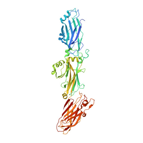
    The Streptococcus mutans antigen I/II (AgI/II) is a cell surface-localized protein that adheres to salivary components and extracellular matrix molecules. Here we report the 2.5 Å resolution crystal structure of the complete C-terminal region of AgI/II. The C-terminal region is comprised of three major domains: C(1), C(2), and C(3). Each domain adopts a DE-variant IgG fold, with two β-sheets whose A and F strands are linked through an intramolecular isopeptide bond. The adherence of the C-terminal AgI/II fragments to the putative tooth surface receptor salivary agglutinin (SAG), as monitored by surface plasmon resonance, indicated that the minimal region of binding was contained within the first and second DE-variant-IgG domains (C(1) and C(2)) of the C terminus. The minimal C-terminal region that could inhibit S. mutans adherence to SAG was also confirmed to be within the C(1) and C(2) domains. Competition experiments demonstrated that the C- and N-terminal regions of AgI/II adhere to distinct sites on SAG. A cleft formed at the intersection between these C(1) and C(2) domains bound glucose molecules from the cryo-protectant solution, revealing a putative binding site for its highly glycosylated receptor SAG. Finally, electron microscopy images confirmed the elongated structure of AgI/II and enabled building a composite tertiary model that encompasses its two distinct binding regions.


  • Organizational Affiliation

    Department of Physiology and Biophysics, University of Alabama, Birmingham, Alabama 35294, USA.


Macromolecules
Find similar proteins by:  (by identity cutoff)  |  3D Structure
Entity ID: 1
MoleculeChains Sequence LengthOrganismDetailsImage
Major cell-surface adhesin PAc
A, B
516Streptococcus mutansMutation(s): 0 
Gene Names: pac
UniProt
Find proteins for P11657 (Streptococcus mutans)
Explore P11657 
Go to UniProtKB:  P11657
Entity Groups  
Sequence Clusters30% Identity50% Identity70% Identity90% Identity95% Identity100% Identity
UniProt GroupP11657
Sequence Annotations
Expand
  • Reference Sequence
Small Molecules
Ligands 3 Unique
IDChains Name / Formula / InChI Key2D Diagram3D Interactions
GLC
Query on GLC

Download Ideal Coordinates CCD File 
G [auth A]
H [auth A]
I [auth A]
J [auth A]
K [auth A]
G [auth A],
H [auth A],
I [auth A],
J [auth A],
K [auth A],
O [auth B],
P [auth B],
Q [auth B]
alpha-D-glucopyranose
C6 H12 O6
WQZGKKKJIJFFOK-DVKNGEFBSA-N
CA
Query on CA

Download Ideal Coordinates CCD File 
C [auth A]
D [auth A]
E [auth A]
L [auth B]
M [auth B]
C [auth A],
D [auth A],
E [auth A],
L [auth B],
M [auth B],
N [auth B]
CALCIUM ION
Ca
BHPQYMZQTOCNFJ-UHFFFAOYSA-N
MG
Query on MG

Download Ideal Coordinates CCD File 
F [auth A]MAGNESIUM ION
Mg
JLVVSXFLKOJNIY-UHFFFAOYSA-N
Experimental Data & Validation

Experimental Data

  • Method: X-RAY DIFFRACTION
  • Resolution: 2.50 Å
  • R-Value Free: 0.244 
  • R-Value Work: 0.208 
  • R-Value Observed: 0.208 
  • Space Group: I 2 2 2
Unit Cell:
Length ( Å )Angle ( ˚ )
a = 108.495α = 90
b = 156.36β = 90
c = 213.648γ = 90
Software Package:
Software NamePurpose
HKL-2000data collection
SHARPphasing
CNSrefinement
HKL-2000data reduction
HKL-2000data scaling

Structure Validation

View Full Validation Report


Ligand Structure Quality Assessment 


Entry History 

Deposition Data

Revision History  (Full details and data files)

  • Version 1.0: 2011-04-20
    Type: Initial release
  • Version 1.1: 2011-07-13
    Changes: Version format compliance
  • Version 1.2: 2020-07-29
    Type: Remediation
    Reason: Carbohydrate remediation
    Changes: Data collection, Database references, Derived calculations, Structure summary