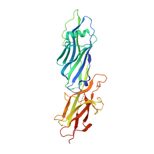
Two autonomous structural modules in the fimbrial shaft adhesin FimA mediate Actinomyces interactions with streptococci and host cells during oral biofilm development.
Mishra, A., Devarajan, B., Reardon, M.E., Dwivedi, P., Krishnan, V., Cisar, J.O., Das, A., Narayana, S.V., Ton-That, H.(2011) Mol Microbiol 81: 1205-1220
- PubMed: 21696465
- DOI: https://doi.org/10.1111/j.1365-2958.2011.07745.x
- Primary Citation of Related Structures:
3QDH - PubMed Abstract:
By combining X-ray crystallography and modelling, we describe here the atomic structure of distinct adhesive moieties of FimA, the shaft fimbrillin of Actinomyces type 2 fimbriae, which uniquely mediates the receptor-dependent intercellular interactions between Actinomyces and oral streptococci as well as host cells during the development of oral biofilms. The FimA adhesin is built with three IgG-like domains, each of which harbours an intramolecular isopeptide bond, previously described in several Gram-positive pilins. Genetic and biochemical studies demonstrate that although these isopeptide bonds are dispensable for fimbrial assembly, cell-cell interactions and biofilm formation, they contribute significantly to the proteolytic stability of FimA. Remarkably, FimA harbours two autonomous adhesive modules, which structurally resemble the Staphylococcus aureus Cna B domain. Each isolated module can bind the plasma glycoprotein asialofetuin as well as the polysaccharide receptors present on the surface of oral streptococci and epithelial cells. Thus, FimA should serve as an excellent paradigm for the development of therapeutic strategies and elucidating the precise molecular mechanisms underlying the interactions between cellular receptors and Gram-positive fimbriae.
Organizational Affiliation:
Department of Microbiology & Molecular Genetics, University of Texas Health Science Center, Houston, TX, USA.