3PIE

Crystal structure of the 5'->3' exoribonuclease Xrn1, E178Q mutant


Experimental Data Snapshot

  • Method: X-RAY DIFFRACTION
  • Resolution: 2.90 Å
  • R-Value Free: 0.305 
  • R-Value Work: 0.241 
  • R-Value Observed: 0.244 

wwPDB Validation   3D Report Full Report


This is version 1.4 of the entry. See complete history


Literature

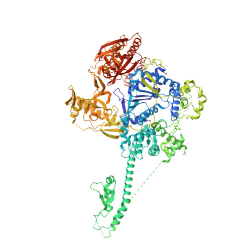
Structural and biochemical studies of the 5' -> 3' exoribonuclease Xrn1.

Chang, J.H.Xiang, S.Xiang, K.Manley, J.L.Tong, L.

(2011) Nat Struct Mol Biol 18: 270-276

  • DOI: https://doi.org/10.1038/nsmb.1984
  • Primary Citation of Related Structures:  
    3PIE, 3PIF

  • PubMed Abstract: 

    The 5'→3' exoribonucleases (XRNs) have important functions in transcription, RNA metabolism and RNA interference. The structure of Rat1 (also known as Xrn2) showed that the two highly conserved regions of XRNs form a single, large domain that defines the active site of the enzyme. Xrn1 has a 510-residue segment after the conserved regions that is required for activity but is absent from Rat1/Xrn2. Here we report the crystal structures of Kluyveromyces lactis Xrn1 (residues 1-1,245, E178Q mutant), alone and in complex with a Mn(2+) ion in the active site. The 510-residue segment contains four domains (D1-D4), located far from the active site. Our mutagenesis and biochemical studies show that their functional importance results from their ability to stabilize the conformation of the N-terminal segment of Xrn1. These domains might also constitute a platform that interacts with protein partners of Xrn1.


  • Organizational Affiliation

    Department of Biological Sciences, Columbia University, New York, NY, USA.


Macromolecules
Find similar proteins by:  (by identity cutoff)  |  3D Structure
Entity ID: 1
MoleculeChains Sequence LengthOrganismDetailsImage
5'->3' EXORIBONUCLEASE (Xrn1)
A, B, C, D
1,155Kluyveromyces lactisMutation(s): 1 
Gene Names: KLLA0F22385g
UniProt
Find proteins for Q6CJ09 (Kluyveromyces lactis (strain ATCC 8585 / CBS 2359 / DSM 70799 / NBRC 1267 / NRRL Y-1140 / WM37))
Explore Q6CJ09 
Go to UniProtKB:  Q6CJ09
Entity Groups  
Sequence Clusters30% Identity50% Identity70% Identity90% Identity95% Identity100% Identity
UniProt GroupQ6CJ09
Sequence Annotations
Expand
  • Reference Sequence
Experimental Data & Validation

Experimental Data

  • Method: X-RAY DIFFRACTION
  • Resolution: 2.90 Å
  • R-Value Free: 0.305 
  • R-Value Work: 0.241 
  • R-Value Observed: 0.244 
  • Space Group: P 1
Unit Cell:
Length ( Å )Angle ( ˚ )
a = 115.72α = 110.07
b = 132.289β = 105.7
c = 143.89γ = 103.75
Software Package:
Software NamePurpose
HKL-2000data collection
PHASERphasing
REFMACrefinement
HKL-2000data reduction
SCALEPACKdata scaling

Structure Validation

View Full Validation Report


Entry History 

Deposition Data

Revision History  (Full details and data files)

  • Version 1.0: 2011-02-09
    Type: Initial release
  • Version 1.1: 2011-07-13
    Changes: Version format compliance
  • Version 1.2: 2013-06-19
    Changes: Database references
  • Version 1.3: 2017-08-02
    Changes: Refinement description, Source and taxonomy
  • Version 1.4: 2024-02-21
    Changes: Data collection, Database references