3PFF

Truncated human atp-citrate lyase with ADP and tartrate bound


Experimental Data Snapshot

  • Method: X-RAY DIFFRACTION
  • Resolution: 2.30 Å
  • R-Value Free: 0.228 
  • R-Value Work: 0.168 
  • R-Value Observed: 0.171 

wwPDB Validation   3D Report Full Report


Ligand Structure Quality Assessment 


This is version 1.3 of the entry. See complete history


Literature

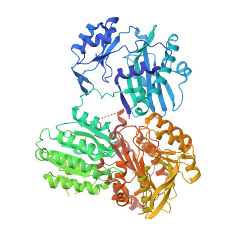
ADP-Mg2+ bound to the ATP-grasp domain of ATP-citrate lyase.

Sun, T.Hayakawa, K.Fraser, M.E.

(2011) Acta Crystallogr Sect F Struct Biol Cryst Commun 67: 1168-1172

  • DOI: https://doi.org/10.1107/S1744309111028363
  • Primary Citation of Related Structures:  
    3PFF

  • PubMed Abstract: 

    Human ATP-citrate lyase (EC 2.3.3.8) is the cytoplasmic enzyme that catalyzes the production of acetyl-CoA from citrate, CoA and ATP. The amino-terminal portion of the enzyme, containing residues 1-817, was crystallized in the presence of tartrate, ATP and magnesium ions. The crystals diffracted to 2.3 Å resolution. The structure shows ADP-Mg(2+) bound to the domain that possesses the ATP-grasp fold. The structure demonstrates that this crystal form could be used to investigate the structures of complexes with inhibitors of ATP-citrate lyase that bind at either the citrate- or ATP-binding site.


  • Organizational Affiliation

    Department of Biological Sciences, University of Calgary, 2500 University Drive NW, Calgary, AB T2N 1N4, Canada.


Macromolecules
Find similar proteins by:  (by identity cutoff)  |  3D Structure
Entity ID: 1
MoleculeChains Sequence LengthOrganismDetailsImage
ATP-citrate synthase829Homo sapiensMutation(s): 0 
Gene Names: ACLY
EC: 2.3.3.8
UniProt & NIH Common Fund Data Resources
Find proteins for P53396 (Homo sapiens)
Explore P53396 
Go to UniProtKB:  P53396
PHAROS:  P53396
GTEx:  ENSG00000131473 
Entity Groups  
Sequence Clusters30% Identity50% Identity70% Identity90% Identity95% Identity100% Identity
UniProt GroupP53396
Sequence Annotations
Expand
  • Reference Sequence
Experimental Data & Validation

Experimental Data

  • Method: X-RAY DIFFRACTION
  • Resolution: 2.30 Å
  • R-Value Free: 0.228 
  • R-Value Work: 0.168 
  • R-Value Observed: 0.171 
  • Space Group: C 1 2 1
Unit Cell:
Length ( Å )Angle ( ˚ )
a = 167.54α = 90
b = 61.7β = 125.47
c = 107.98γ = 90
Software Package:
Software NamePurpose
MxDCdata collection
PHENIXmodel building
PHENIXrefinement
MOSFLMdata reduction
SCALAdata scaling
PHENIXphasing

Structure Validation

View Full Validation Report


Ligand Structure Quality Assessment 


Entry History 

Deposition Data

Revision History  (Full details and data files)

  • Version 1.0: 2011-10-12
    Type: Initial release
  • Version 1.1: 2012-04-11
    Changes: Database references
  • Version 1.2: 2017-11-08
    Changes: Refinement description
  • Version 1.3: 2023-09-06
    Changes: Data collection, Database references, Derived calculations, Refinement description