3OMJ

Structural Basis for Cyclic Py-Im Polyamide Allosteric Inhibition of Nuclear Receptor Binding

  • Classification: DNA
  • Mutation(s): No 

  • Deposited: 2010-08-27 Released: 2010-09-08 
  • Deposition Author(s): Chenoweth, D.M.

Experimental Data Snapshot

  • Method: X-RAY DIFFRACTION
  • Resolution: 0.95 Å
  • R-Value Free: 0.124 
  • R-Value Work: 0.112 
  • R-Value Observed: 0.113 

wwPDB Validation   3D Report Full Report


Ligand Structure Quality Assessment 


This is version 1.2 of the entry. See complete history


Literature

Structural basis for cyclic py-im polyamide allosteric inhibition of nuclear receptor binding.

Chenoweth, D.M.Dervan, P.B.

(2010) J Am Chem Soc 132: 14521-14529

  • DOI: https://doi.org/10.1021/ja105068b
  • Primary Citation of Related Structures:  
    3OMJ

  • PubMed Abstract: 

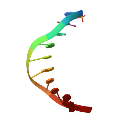
    Pyrrole-imidazole polyamides are a class of small molecules that can be programmed to bind a broad repertoire of DNA sequences, disrupt transcription factor-DNA interfaces, and modulate gene expression pathways in cell culture experiments. In this paper we describe a high-resolution X-ray crystal structure of a β-amino turn-linked eight-ring cyclic Py-Im polyamide bound to the central six base pairs of the sequence d(5'-CCAGTACTGG-3')(2), revealing significant modulation of DNA shape. We compare the DNA structural perturbations induced by DNA-binding transcripton factors, androgen receptor and glucocorticoid receptor, in the major groove to those induced by cyclic polyamide binding in the minor groove. The cyclic polyamide is an allosteric modulator that perturbs the DNA structure in such a way that nuclear receptor protein binding is no longer compatible. This allosteric perturbation of the DNA helix provides a molecular basis for disruption of transcription factor-DNA interfaces by small molecules, a minimum step in chemical control of gene networks.


  • Organizational Affiliation

    Division of Chemistry and Chemical Engineering, California Institute of Technology, Pasadena, California 91125, USA.


Macromolecules

Find similar nucleic acids by:  Sequence   |   3D Structure  

Entity ID: 1
MoleculeChains LengthOrganismImage
5'-D(*CP*(C38)P*AP*GP*TP*AP*CP*TP*GP*G)-3'
A, B
10N/A
Sequence Annotations
Expand
  • Reference Sequence
Small Molecules
Ligands 2 Unique
IDChains Name / Formula / InChI Key2D Diagram3D Interactions
1P2
Query on 1P2

Download Ideal Coordinates CCD File 
F [auth B](23R,52R)-23,52-diamino-5,11,17,28,34,40,46,57-octamethyl-2,5,8,11,14,17,20,25,28,31,34,37,40,43,46,49,54,57,60,64-icosaazanonacyclo[54.2.1.1~4,7~.1~10,13~.1~16,19~.1~27,30~.1~33,36~.1~39,42~.1~45,48~]hexahexaconta-1(58),4(66),6,10(65),12,16(64),18,27(63),29,33(62),35,39(61),41,45(60),47,56(59)-hexadecaene-3,9,15,21,26,32,38,44,50,55-decone
C54 H62 N22 O10
KELPAVUCWDTHOZ-VSGBNLITSA-N
CA
Query on CA

Download Ideal Coordinates CCD File 
C [auth A],
D [auth A],
E [auth A],
G [auth B]
CALCIUM ION
Ca
BHPQYMZQTOCNFJ-UHFFFAOYSA-N
Experimental Data & Validation

Experimental Data

  • Method: X-RAY DIFFRACTION
  • Resolution: 0.95 Å
  • R-Value Free: 0.124 
  • R-Value Work: 0.112 
  • R-Value Observed: 0.113 
  • Space Group: P 41 21 2
Unit Cell:
Length ( Å )Angle ( ˚ )
a = 39.831α = 90
b = 39.831β = 90
c = 84.567γ = 90
Software Package:
Software NamePurpose
CrystalCleardata collection
SHELXSphasing
REFMACrefinement
MOSFLMdata reduction
SCALAdata scaling

Structure Validation

View Full Validation Report


Ligand Structure Quality Assessment 


Entry History 

Deposition Data

Revision History  (Full details and data files)

  • Version 1.0: 2010-09-08
    Type: Initial release
  • Version 1.1: 2011-07-13
    Changes: Version format compliance
  • Version 1.2: 2024-02-21
    Changes: Advisory, Data collection, Database references, Derived calculations