3OK8

I-BAR OF PinkBAR


Experimental Data Snapshot

  • Method: X-RAY DIFFRACTION
  • Resolution: 2.25 Å
  • R-Value Free: 0.279 
  • R-Value Work: 0.221 
  • R-Value Observed: 0.225 

wwPDB Validation   3D Report Full Report


This is version 1.3 of the entry. See complete history


Literature

Pinkbar is an epithelial-specific BAR domain protein that generates planar membrane structures.

Pykalainen, A.Boczkowska, M.Zhao, H.Saarikangas, J.Rebowski, G.Jansen, M.Hakanen, J.Koskela, E.V.Peranen, J.Vihinen, H.Jokitalo, E.Salminen, M.Ikonen, E.Dominguez, R.Lappalainen, P.

(2011) Nat Struct Mol Biol 18: 902-907

  • DOI: https://doi.org/10.1038/nsmb.2079
  • Primary Citation of Related Structures:  
    3OK8

  • PubMed Abstract: 

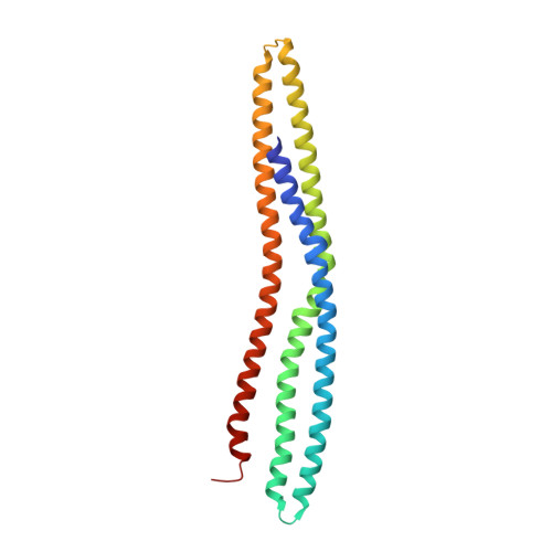
    Bin/amphipysin/Rvs (BAR)-domain proteins sculpt cellular membranes and have key roles in processes such as endocytosis, cell motility and morphogenesis. BAR domains are divided into three subfamilies: BAR- and F-BAR-domain proteins generate positive membrane curvature and stabilize cellular invaginations, whereas I-BAR-domain proteins induce negative curvature and stabilize protrusions. We show that a previously uncharacterized member of the I-BAR subfamily, Pinkbar, is specifically expressed in intestinal epithelial cells, where it localizes to Rab13-positive vesicles and to the plasma membrane at intercellular junctions. Notably, the BAR domain of Pinkbar does not induce membrane tubulation but promotes the formation of planar membrane sheets. Structural and mutagenesis analyses reveal that the BAR domain of Pinkbar has a relatively flat lipid-binding interface and that it assembles into sheet-like oligomers in crystals and in solution, which may explain its unique membrane-deforming activity.


  • Organizational Affiliation

    Institute of Biotechnology, University of Helsinki, Helsinki, Finland.


Macromolecules
Find similar proteins by:  (by identity cutoff)  |  3D Structure
Entity ID: 1
MoleculeChains Sequence LengthOrganismDetailsImage
Brain-specific angiogenesis inhibitor 1-associated protein 2-like protein 2
A, B
222Mus musculusMutation(s): 0 
Gene Names: Baiap2l2PinkBAR
UniProt & NIH Common Fund Data Resources
Find proteins for Q80Y61 (Mus musculus)
Explore Q80Y61 
Go to UniProtKB:  Q80Y61
IMPC:  MGI:2652819
Entity Groups  
Sequence Clusters30% Identity50% Identity70% Identity90% Identity95% Identity100% Identity
UniProt GroupQ80Y61
Sequence Annotations
Expand
  • Reference Sequence
Small Molecules
Ligands 1 Unique
IDChains Name / Formula / InChI Key2D Diagram3D Interactions
GOL
Query on GOL

Download Ideal Coordinates CCD File 
C [auth A]GLYCEROL
C3 H8 O3
PEDCQBHIVMGVHV-UHFFFAOYSA-N
Experimental Data & Validation

Experimental Data

  • Method: X-RAY DIFFRACTION
  • Resolution: 2.25 Å
  • R-Value Free: 0.279 
  • R-Value Work: 0.221 
  • R-Value Observed: 0.225 
  • Space Group: C 1 2 1
Unit Cell:
Length ( Å )Angle ( ˚ )
a = 99.92α = 90
b = 39.913β = 94.82
c = 120.702γ = 90
Software Package:
Software NamePurpose
CCP4model building
PHENIXrefinement
HKL-2000data reduction
HKL-2000data scaling
CCP4phasing

Structure Validation

View Full Validation Report


Entry History 

Deposition Data

Revision History  (Full details and data files)

  • Version 1.0: 2011-07-13
    Type: Initial release
  • Version 1.1: 2011-07-20
    Changes: Database references
  • Version 1.2: 2011-08-17
    Changes: Database references
  • Version 1.3: 2024-02-21
    Changes: Data collection, Database references, Derived calculations