3OJL

Native structure of the UDP-N-acetyl-mannosamine dehydrogenase Cap5O from Staphylococcus aureus


Experimental Data Snapshot

  • Method: X-RAY DIFFRACTION
  • Resolution: 2.80 Å
  • R-Value Free: 0.263 
  • R-Value Work: 0.217 
  • R-Value Observed: 0.219 

wwPDB Validation   3D Report Full Report


Ligand Structure Quality Assessment 


This is version 1.2 of the entry. See complete history


Literature

Structure Analysis of the Staphylococcus aureus UDP-N-acetyl-mannosamine Dehydrogenase Cap5O Involved in Capsular Polysaccharide Biosynthesis.

Gruszczyk, J.Fleurie, A.Olivares-Illana, V.Bechet, E.Zanella-Cleon, I.Morera, S.Meyer, P.Pompidor, G.Kahn, R.Grangeasse, C.Nessler, S.

(2011) J Biol Chem 286: 17112-17121

  • DOI: https://doi.org/10.1074/jbc.M110.216002
  • Primary Citation of Related Structures:  
    3OJL, 3OJO

  • PubMed Abstract: 

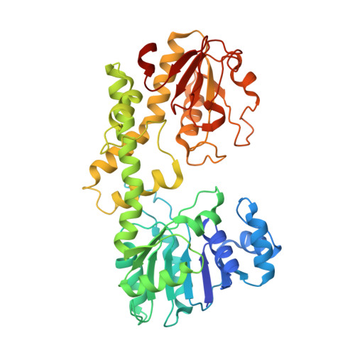
    Bacterial UDP-sugar dehydrogenases are part of the biosynthesis pathway of extracellular polysaccharides. These compounds act as important virulence factors by protecting the cell from opsonophagocytosis and complement-mediated killing. In Staphylococcus aureus, the protein Cap5O catalyzes the oxidation of UDP-N-acetyl-mannosamine to UDP-N-acetyl-mannosaminuronic acid. Cap5O is crucial for the production of serotype 5 capsular polysaccharide that prevents the interaction of bacteria with both phagocytic and nonphagocytic eukaryotic cells. However, details of its catalytic mechanism remain unknown. We thus crystallized Cap5O and solved the first structure of an UDP-N-acetyl-mannosamine dehydrogenase. This study revealed that the catalytic cysteine makes a disulfide bond that has never been observed in other structurally characterized members of the NDP-sugar dehydrogenase family. Biochemical and mutagenesis experiments demonstrated that the formation of this disulfide bridge regulates the activity of Cap5O. We also identified two arginine residues essential for Cap5O activity. Previous data suggested that Cap5O is activated by tyrosine phosphorylation, so we characterized the phosphorylation site and examined the underlying regulatory mechanism.


  • Organizational Affiliation

    Laboratoire d'Enzymologie et Biochimie Structurales, Centre de Recherche de Gif, CNRS, 91198 Gif sur Yvette, France.


Macromolecules
Find similar proteins by:  (by identity cutoff)  |  3D Structure
Entity ID: 1
MoleculeChains Sequence LengthOrganismDetailsImage
Cap5O
A, B
431Staphylococcus aureusMutation(s): 0 
Gene Names: cap5O
UniProt
Find proteins for P95708 (Staphylococcus aureus)
Explore P95708 
Go to UniProtKB:  P95708
Entity Groups  
Sequence Clusters30% Identity50% Identity70% Identity90% Identity95% Identity100% Identity
UniProt GroupP95708
Sequence Annotations
Expand
  • Reference Sequence
Small Molecules
Ligands 1 Unique
IDChains Name / Formula / InChI Key2D Diagram3D Interactions
NAD
Query on NAD

Download Ideal Coordinates CCD File 
C [auth A],
D [auth B]
NICOTINAMIDE-ADENINE-DINUCLEOTIDE
C21 H27 N7 O14 P2
BAWFJGJZGIEFAR-NNYOXOHSSA-N
Experimental Data & Validation

Experimental Data

  • Method: X-RAY DIFFRACTION
  • Resolution: 2.80 Å
  • R-Value Free: 0.263 
  • R-Value Work: 0.217 
  • R-Value Observed: 0.219 
  • Space Group: P 21 21 21
Unit Cell:
Length ( Å )Angle ( ˚ )
a = 77.901α = 90
b = 87.264β = 90
c = 131.576γ = 90
Software Package:
Software NamePurpose
ADSCdata collection
PHASERphasing
BUSTERrefinement
MOSFLMdata reduction
SCALAdata scaling

Structure Validation

View Full Validation Report


Ligand Structure Quality Assessment 


Entry History 

Deposition Data

Revision History  (Full details and data files)

  • Version 1.0: 2011-03-23
    Type: Initial release
  • Version 1.1: 2011-07-13
    Changes: Version format compliance
  • Version 1.2: 2023-09-06
    Changes: Data collection, Database references, Derived calculations, Refinement description