3O6G

Ligand-binding domain of GluA2 (flip) ionotropic glutamate receptor in complex with an allosteric modulator


Experimental Data Snapshot

  • Method: X-RAY DIFFRACTION
  • Resolution: 1.80 Å
  • R-Value Free: 0.223 
  • R-Value Work: 0.189 
  • R-Value Observed: 0.191 

wwPDB Validation   3D Report Full Report


Ligand Structure Quality Assessment 


This is version 1.2 of the entry. See complete history


Literature

A novel series of positive modulators of the AMPA receptor: structure-based lead optimization.

Jamieson, C.Campbell, R.A.Cumming, I.A.Gillen, K.J.Gillespie, J.Kazemier, B.Kiczun, M.Lamont, Y.Lyons, A.J.Maclean, J.K.Martin, F.Moir, E.M.Morrow, J.A.Pantling, J.Rankovic, Z.Smith, L.

(2010) Bioorg Med Chem Lett 20: 6072-6075

  • DOI: https://doi.org/10.1016/j.bmcl.2010.08.063
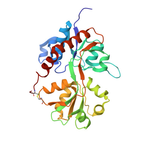
  • Primary Citation of Related Structures:  
    3O6G, 3O6H, 3O6I

  • PubMed Abstract: 

    Starting from lead compound 1, we demonstrate how X-ray structural data can be used to understand SAR and expediently optimize bioavailability in a novel series of AMPA receptor modulators, furnishing 5 with improved bioavailability and robust in vivo activity.


  • Organizational Affiliation

    Merck Research Laboratories, MSD, Newhouse, Motherwell, Lanarkshire, UK. craig.jamieson@strath.ac.uk


Macromolecules
Find similar proteins by:  (by identity cutoff)  |  3D Structure
Entity ID: 1
MoleculeChains Sequence LengthOrganismDetailsImage
Glutamate receptor 2263Rattus norvegicusMutation(s): 0 
Gene Names: Gria2Glur2
UniProt
Find proteins for P19491 (Rattus norvegicus)
Explore P19491 
Go to UniProtKB:  P19491
Entity Groups  
Sequence Clusters30% Identity50% Identity70% Identity90% Identity95% Identity100% Identity
UniProt GroupP19491
Sequence Annotations
Expand
  • Reference Sequence
Small Molecules
Ligands 4 Unique
IDChains Name / Formula / InChI Key2D Diagram3D Interactions
O27
Query on O27

Download Ideal Coordinates CCD File 
F [auth A]N-[(3R)-pyrrolidin-3-yl]-2-({[3-(trifluoromethyl)-4,5,6,7-tetrahydro-1H-indazol-1-yl]acetyl}amino)-4,5,6,7-tetrahydro-1-benzothiophene-3-carboxamide
C23 H28 F3 N5 O2 S
CFYCEUGQUDABFL-CYBMUJFWSA-N
GLU
Query on GLU

Download Ideal Coordinates CCD File 
B [auth A]GLUTAMIC ACID
C5 H9 N O4
WHUUTDBJXJRKMK-VKHMYHEASA-N
SO4
Query on SO4

Download Ideal Coordinates CCD File 
C [auth A],
D [auth A],
E [auth A]
SULFATE ION
O4 S
QAOWNCQODCNURD-UHFFFAOYSA-L
EDO
Query on EDO

Download Ideal Coordinates CCD File 
G [auth A],
H [auth A],
I [auth A],
J [auth A]
1,2-ETHANEDIOL
C2 H6 O2
LYCAIKOWRPUZTN-UHFFFAOYSA-N
Experimental Data & Validation

Experimental Data

  • Method: X-RAY DIFFRACTION
  • Resolution: 1.80 Å
  • R-Value Free: 0.223 
  • R-Value Work: 0.189 
  • R-Value Observed: 0.191 
  • Space Group: P 21 21 2
Unit Cell:
Length ( Å )Angle ( ˚ )
a = 64.09α = 90
b = 87.18β = 90
c = 47.32γ = 90
Software Package:
Software NamePurpose
CrystalCleardata collection
AMoREphasing
REFMACrefinement
CrystalCleardata reduction
CrystalCleardata scaling

Structure Validation

View Full Validation Report


Ligand Structure Quality Assessment 


Entry History 

Revision History  (Full details and data files)

  • Version 1.0: 2010-09-15
    Type: Initial release
  • Version 1.1: 2011-07-13
    Changes: Version format compliance
  • Version 1.2: 2017-08-09
    Changes: Refinement description, Source and taxonomy