3O39

Crystal Structure of SPY


Experimental Data Snapshot

  • Method: X-RAY DIFFRACTION
  • Resolution: 2.60 Å
  • R-Value Free: 0.281 
  • R-Value Work: 0.243 
  • R-Value Observed: 0.245 

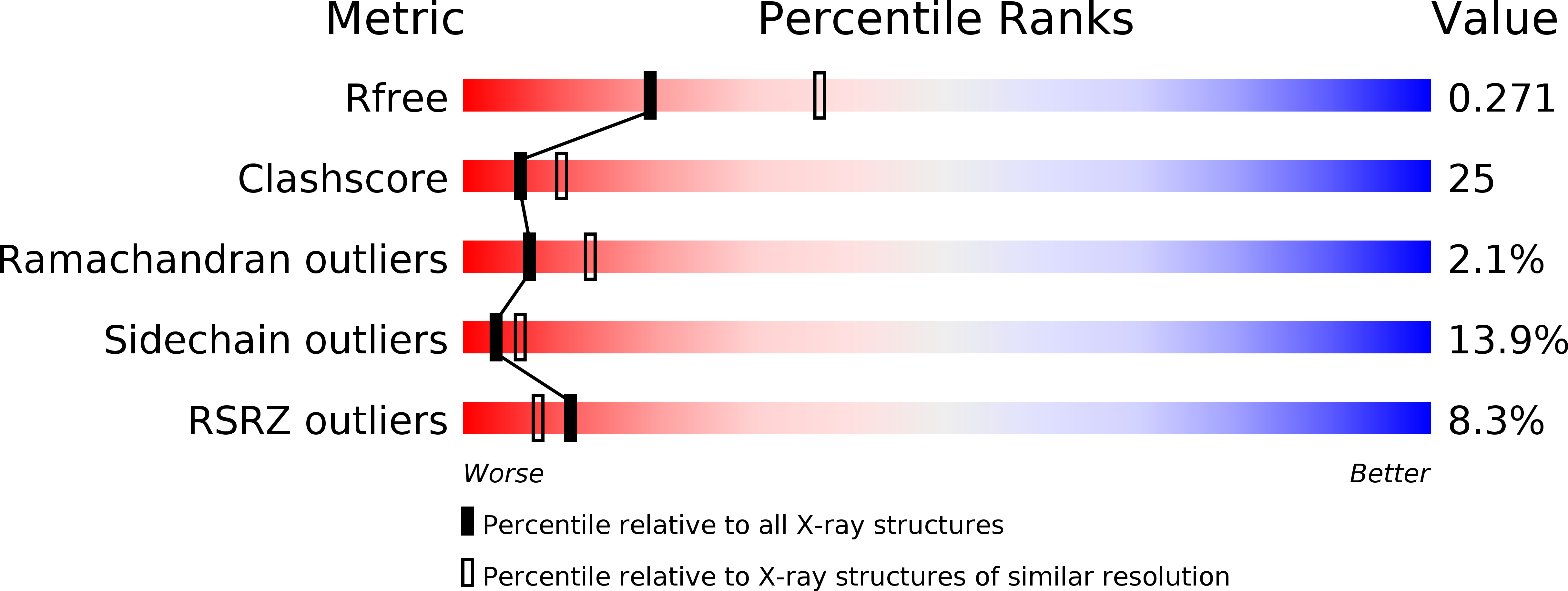
wwPDB Validation   3D Report Full Report


This is version 1.3 of the entry. See complete history


Literature

Genetic selection designed to stabilize proteins uncovers a chaperone called Spy.

Quan, S.Koldewey, P.Tapley, T.Kirsch, N.Ruane, K.M.Pfizenmaier, J.Shi, R.Hofmann, S.Foit, L.Ren, G.Jakob, U.Xu, Z.Cygler, M.Bardwell, J.C.

(2011) Nat Struct Mol Biol 18: 262-269

  • DOI: https://doi.org/10.1038/nsmb.2016
  • Primary Citation of Related Structures:  
    3O39

  • PubMed Abstract: 

    To optimize the in vivo folding of proteins, we linked protein stability to antibiotic resistance, thereby forcing bacteria to effectively fold and stabilize proteins. When we challenged Escherichia coli to stabilize a very unstable periplasmic protein, it massively overproduced a periplasmic protein called Spy, which increases the steady-state levels of a set of unstable protein mutants up to 700-fold. In vitro studies demonstrate that the Spy protein is an effective ATP-independent chaperone that suppresses protein aggregation and aids protein refolding. Our strategy opens up new routes for chaperone discovery and the custom tailoring of the in vivo folding environment. Spy forms thin, apparently flexible cradle-shaped dimers. The structure of Spy is unlike that of any previously solved chaperone, making it the prototypical member of a new class of small chaperones that facilitate protein refolding in the absence of energy cofactors.


  • Organizational Affiliation

    Howard Hughes Medical Institute, Chevy Chase, MD, USA.


Macromolecules
Find similar proteins by:  (by identity cutoff)  |  3D Structure
Entity ID: 1
MoleculeChains Sequence LengthOrganismDetailsImage
Periplasmic protein related to spheroblast formation
A, B
108Escherichia coli O157:H7 str. EDL933Mutation(s): 0 
Gene Names: ECs2449spyZ2775
UniProt
Find proteins for Q8XDZ4 (Escherichia coli O157:H7)
Explore Q8XDZ4 
Go to UniProtKB:  Q8XDZ4
Entity Groups  
Sequence Clusters30% Identity50% Identity70% Identity90% Identity95% Identity100% Identity
UniProt GroupQ8XDZ4
Sequence Annotations
Expand
  • Reference Sequence
Experimental Data & Validation

Experimental Data

  • Method: X-RAY DIFFRACTION
  • Resolution: 2.60 Å
  • R-Value Free: 0.281 
  • R-Value Work: 0.243 
  • R-Value Observed: 0.245 
  • Space Group: P 62
Unit Cell:
Length ( Å )Angle ( ˚ )
a = 68.971α = 90
b = 68.971β = 90
c = 124.281γ = 120
Software Package:
Software NamePurpose
PHENIXrefinement
SCALEPACKdata scaling
SHARPphasing
RESOLVEphasing
REFMACrefinement
PDB_EXTRACTdata extraction
DENZOdata reduction

Structure Validation

View Full Validation Report


Entry History 

Deposition Data

Revision History  (Full details and data files)

  • Version 1.0: 2011-02-16
    Type: Initial release
  • Version 1.1: 2011-07-13
    Changes: Version format compliance
  • Version 1.2: 2017-11-08
    Changes: Refinement description
  • Version 1.3: 2019-07-17
    Changes: Data collection, Derived calculations, Refinement description