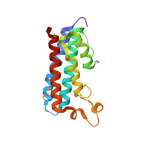
3O10

Crystal structure of the HEPN domain from human sacsin


Experimental Data Snapshot

  • Method: X-RAY DIFFRACTION
  • Resolution: 1.90 Å
  • R-Value Free: 0.225 
  • R-Value Work: 0.184 
  • R-Value Observed: 0.186 

wwPDB Validation   3D Report Full Report


This is version 1.2 of the entry. See complete history


Literature

Structural basis of defects in the sacsin HEPN domain responsible for autosomal recessive spastic ataxia of Charlevoix-Saguenay (ARSACS).

Kozlov, G.Denisov, A.Y.Girard, M.Dicaire, M.J.Hamlin, J.McPherson, P.S.Brais, B.Gehring, K.

(2011) J Biol Chem 286: 20407-20412

  • DOI: https://doi.org/10.1074/jbc.M111.232884
  • Primary Citation of Related Structures:  
    3O10

  • PubMed Abstract: 

    Sacsin is a 520-kDa protein mutated in the early-onset neurodevelopmental and neurodegenerative disease autosomal recessive spastic ataxia of Charlevoix-Saguenay (ARSACS). The C terminus of the protein contains an HEPN (higher eukaryotes and prokaryotes nucleotide-binding) domain of unknown function. Here, we determined the high-resolution 1.9-Å crystal structure of the HEPN domain from human sacsin. The structure is composed of five parallel α-helices with a large loop of several short helical segments. Two HEPN protomers assemble as a dimer to form a large positively charged cavity at the dimer interface that binds GTP and other nucleotides. The crystal structure reveals that the ARSACS N4549D mutation disrupts dimerization and protein folding. This study provides novel insights into the oligomerization state of sacsin and functions that are lost in mutations that cause ARSACS.


  • Organizational Affiliation

    Department of Biochemistry, Groupe de Recherche Axé sur la Structure des Protéines, McGill University, Montréal, Québec H3G 0B1, Canada.


Macromolecules
Find similar proteins by:  (by identity cutoff)  |  3D Structure
Entity ID: 1
MoleculeChains Sequence LengthOrganismDetailsImage
Sacsin
A, B, C, D
141Homo sapiensMutation(s): 0 
Gene Names: SACSKIAA0730
UniProt & NIH Common Fund Data Resources
Find proteins for Q9NZJ4 (Homo sapiens)
Explore Q9NZJ4 
Go to UniProtKB:  Q9NZJ4
PHAROS:  Q9NZJ4
GTEx:  ENSG00000151835 
Entity Groups  
Sequence Clusters30% Identity50% Identity70% Identity90% Identity95% Identity100% Identity
UniProt GroupQ9NZJ4
Sequence Annotations
Expand
  • Reference Sequence
Small Molecules
Modified Residues  1 Unique
IDChains TypeFormula2D DiagramParent
MSE
Query on MSE
A, B, C, D
L-PEPTIDE LINKINGC5 H11 N O2 SeMET
Experimental Data & Validation

Experimental Data

  • Method: X-RAY DIFFRACTION
  • Resolution: 1.90 Å
  • R-Value Free: 0.225 
  • R-Value Work: 0.184 
  • R-Value Observed: 0.186 
  • Space Group: P 32 2 1
Unit Cell:
Length ( Å )Angle ( ˚ )
a = 72.581α = 90
b = 72.581β = 90
c = 201.18γ = 120
Software Package:
Software NamePurpose
ADSCdata collection
SHELXSphasing
REFMACrefinement
HKL-2000data reduction
HKL-2000data scaling

Structure Validation

View Full Validation Report


Entry History 

Deposition Data

Revision History  (Full details and data files)

  • Version 1.0: 2011-03-30
    Type: Initial release
  • Version 1.1: 2011-07-13
    Changes: Version format compliance
  • Version 1.2: 2014-07-30
    Changes: Database references