3NPC

Crystal structure of JNK2 complexed with BIRB796


Experimental Data Snapshot

  • Method: X-RAY DIFFRACTION
  • Resolution: 2.35 Å
  • R-Value Free: 0.254 
  • R-Value Work: 0.212 
  • R-Value Observed: 0.214 

wwPDB Validation   3D Report Full Report


Ligand Structure Quality Assessment 


This is version 1.2 of the entry. See complete history


Literature

X-ray crystal structure of JNK2 complexed with the p38alpha inhibitor BIRB796: Insights into the rational design of DFG-out binding MAP kinase inhibitors.

Kuglstatter, A.Ghate, M.Tsing, S.Villasenor, A.G.Shaw, D.Barnett, J.W.Browner, M.F.

(2010) Bioorg Med Chem Lett 20: 5217-5220

  • DOI: https://doi.org/10.1016/j.bmcl.2010.06.157
  • Primary Citation of Related Structures:  
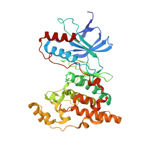
    3NPC

  • PubMed Abstract: 

    JNK2 and p38alpha are closely related mitogen-activated protein kinases that regulate various cellular activities and are considered drug targets for inflammatory diseases. We have determined the X-ray crystal structure of the clinical phase II p38alpha inhibitor BIRB796 bound to its off-target JNK2. This shows for the first time a JNK subfamily member in the DFG-out conformation. The fully resolved activation loop reveals that BIRB796 inhibits JNK2 activation by stabilizing the loop in a position that does not allow its phosphorylation by upstream kinases. The structure suggests that substituents at the BIRB796 morpholino group and modifications of the t-butyl moiety should further increase the p38alpha to JNK2 potency ratio. For the design of selective DFG-out binding JNK2 inhibitors, the binding pocket of the BIRB796 tolyl group may have the best potential.


  • Organizational Affiliation

    Roche Palo Alto, Palo Alto, CA 94304, USA. andreas.kuglstatter@roche.com


Macromolecules
Find similar proteins by:  (by identity cutoff)  |  3D Structure
Entity ID: 1
MoleculeChains Sequence LengthOrganismDetailsImage
Mitogen-activated protein kinase 9
A, B
364Homo sapiensMutation(s): 8 
Gene Names: MAPK9JNK2PRKM9
EC: 2.7.11.24
UniProt & NIH Common Fund Data Resources
Find proteins for P45984 (Homo sapiens)
Explore P45984 
Go to UniProtKB:  P45984
PHAROS:  P45984
GTEx:  ENSG00000050748 
Entity Groups  
Sequence Clusters30% Identity50% Identity70% Identity90% Identity95% Identity100% Identity
UniProt GroupP45984
Sequence Annotations
Expand
  • Reference Sequence
Small Molecules
Ligands 1 Unique
IDChains Name / Formula / InChI Key2D Diagram3D Interactions
B96
Query on B96

Download Ideal Coordinates CCD File 
C [auth A],
D [auth B]
1-(5-TERT-BUTYL-2-P-TOLYL-2H-PYRAZOL-3-YL)-3-[4-(2-MORPHOLIN-4-YL-ETHOXY)-NAPHTHALEN-1-YL]-UREA
C31 H37 N5 O3
MVCOAUNKQVWQHZ-UHFFFAOYSA-N
Binding Affinity Annotations 
IDSourceBinding Affinity
B96 BindingDB:  3NPC Kd: min: 4.6, max: 110 (nM) from 5 assay(s)
IC50: min: 6, max: 1.00e+4 (nM) from 10 assay(s)
PDBBind:  3NPC Kd: 4.6 (nM) from 1 assay(s)
Experimental Data & Validation

Experimental Data

  • Method: X-RAY DIFFRACTION
  • Resolution: 2.35 Å
  • R-Value Free: 0.254 
  • R-Value Work: 0.212 
  • R-Value Observed: 0.214 
  • Space Group: P 21 21 21
Unit Cell:
Length ( Å )Angle ( ˚ )
a = 76.17α = 90
b = 92.339β = 90
c = 112.109γ = 90
Software Package:
Software NamePurpose
ADSCdata collection
PHASERphasing
REFMACrefinement
HKL-2000data reduction
HKL-2000data scaling

Structure Validation

View Full Validation Report


Ligand Structure Quality Assessment 


Entry History 

Deposition Data

Revision History  (Full details and data files)

  • Version 1.0: 2010-08-11
    Type: Initial release
  • Version 1.1: 2011-07-13
    Changes: Version format compliance
  • Version 1.2: 2023-09-06
    Changes: Data collection, Database references, Derived calculations, Refinement description