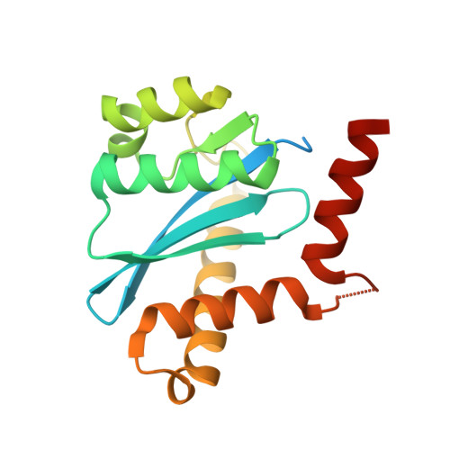
3NF7

Structural basis for a new mechanism of inhibition of HIV integrase identified by fragment screening and structure based design


Experimental Data Snapshot

  • Method: X-RAY DIFFRACTION
  • Resolution: 1.80 Å
  • R-Value Free: 0.207 
  • R-Value Work: 0.162 
  • R-Value Observed: 0.165 

wwPDB Validation   3D Report Full Report


This is version 1.2 of the entry. See complete history


Literature

Structural basis for a new mechanism of inhibition of HIV-1 integrase identified by fragment screening and structure-based design

Rhodes, D.I.Peat, T.S.Vandegraaff, N.Jeevarajah, D.Le, G.Jones, E.D.Smith, J.A.Coates, J.A.Winfield, L.J.Thienthong, N.Newman, J.Lucent, D.Ryan, J.H.Savage, G.P.Francis, C.L.Deadman, J.J.

(2011) Antivir Chem Chemother 21: 155-168

  • DOI: https://doi.org/10.3851/IMP1716
  • Primary Citation of Related Structures:  
    3NF6, 3NF7, 3NF8, 3NF9, 3NFA

  • PubMed Abstract: 

    HIV-1 integrase is a clinically validated therapeutic target for the treatment of HIV-1 infection, with one approved therapeutic currently on the market. This enzyme represents an attractive target for the development of new inhibitors to HIV-1 that are effective against the current resistance mutations.


  • Organizational Affiliation

    Avexa Ltd, Richmond, Australia. drhodes@jdjbioservices.com


Macromolecules
Find similar proteins by:  (by identity cutoff)  |  3D Structure
Entity ID: 1
MoleculeChains Sequence LengthOrganismDetailsImage
Integrase
A, B
183Human immunodeficiency virus 1Mutation(s): 3 
UniProt
Find proteins for P12497 (Human immunodeficiency virus type 1 group M subtype B (isolate NY5))
Explore P12497 
Go to UniProtKB:  P12497
Entity Groups  
Sequence Clusters30% Identity50% Identity70% Identity90% Identity95% Identity100% Identity
UniProt GroupP12497
Sequence Annotations
Expand
  • Reference Sequence
Small Molecules
Ligands 4 Unique
IDChains Name / Formula / InChI Key2D Diagram3D Interactions
CIW
Query on CIW

Download Ideal Coordinates CCD File 
N [auth A],
W [auth B]
5-[(5-chloro-2-oxo-2,3-dihydro-1H-indol-1-yl)methyl]-1,3-benzodioxole-4-carboxylic acid
C17 H12 Cl N O5
ZWUIUQQUOGIKGU-UHFFFAOYSA-N
SO4
Query on SO4

Download Ideal Coordinates CCD File 
C [auth A]
D [auth A]
E [auth A]
F [auth A]
G [auth A]
C [auth A],
D [auth A],
E [auth A],
F [auth A],
G [auth A],
O [auth B],
P [auth B],
Q [auth B],
R [auth B],
S [auth B]
SULFATE ION
O4 S
QAOWNCQODCNURD-UHFFFAOYSA-L
EDO
Query on EDO

Download Ideal Coordinates CCD File 
M [auth A],
V [auth B]
1,2-ETHANEDIOL
C2 H6 O2
LYCAIKOWRPUZTN-UHFFFAOYSA-N
ACY
Query on ACY

Download Ideal Coordinates CCD File 
H [auth A]
I [auth A]
J [auth A]
K [auth A]
L [auth A]
H [auth A],
I [auth A],
J [auth A],
K [auth A],
L [auth A],
T [auth B],
U [auth B]
ACETIC ACID
C2 H4 O2
QTBSBXVTEAMEQO-UHFFFAOYSA-N
Binding Affinity Annotations 
IDSourceBinding Affinity
CIW PDBBind:  3NF7 IC50: 3.40e+5 (nM) from 1 assay(s)
Binding MOAD:  3NF7 IC50: 3.40e+5 (nM) from 1 assay(s)
Experimental Data & Validation

Experimental Data

  • Method: X-RAY DIFFRACTION
  • Resolution: 1.80 Å
  • R-Value Free: 0.207 
  • R-Value Work: 0.162 
  • R-Value Observed: 0.165 
  • Space Group: P 31
Unit Cell:
Length ( Å )Angle ( ˚ )
a = 71.333α = 90
b = 71.333β = 90
c = 66.915γ = 120
Software Package:
Software NamePurpose
Blu-Icedata collection
PHASERphasing
REFMACrefinement
MOSFLMdata reduction
SCALAdata scaling

Structure Validation

View Full Validation Report


Entry History 

Deposition Data

Revision History  (Full details and data files)

  • Version 1.0: 2011-04-27
    Type: Initial release
  • Version 1.1: 2011-07-13
    Changes: Version format compliance
  • Version 1.2: 2012-05-02
    Changes: Database references