Structural basis for branching-enzyme activity of glycoside hydrolase family 57: Structure and stability studies of a novel branching enzyme from the hyperthermophilic archaeon Thermococcus Kodakaraensis KOD1.
Santos, C.R., Tonoli, C.C., Trindade, D.M., Betzel, C., Takata, H., Kuriki, T., Kanai, T., Imanaka, T., Arni, R.K., Murakami, M.T.(2011) Proteins 79: 547-557
- PubMed: 21104698
- DOI: https://doi.org/10.1002/prot.22902
- Primary Citation of Related Structures:
3N8T, 3N92, 3N98 - PubMed Abstract:
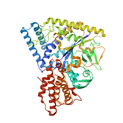
Branching enzymes (BEs) catalyze the formation of branch points in glycogen and amylopectin by cleavage of α-1,4 glycosidic bonds and subsequent transfer to a new α-1,6 position. BEs generally belong to glycoside hydrolase family 13 (GH13); however TK1436, isolated from the hyperthermophilic archaeon Thermococcus kodakaraensis KOD1, is the first GH57 member, which possesses BE activity. To date, the only BE structure that had been determined is a GH13-type from Escherichia coli. Herein, we have determined the crystal structure of TK1436 in the native state and in complex with glucose and substrate mimetics that permitted mapping of the substrate-binding channel and identification of key residues for glucanotransferase activity. Its structure encompasses a distorted (β/α)(7)-barrel juxtaposed to a C-terminal α-helical domain, which also participates in the formation of the active-site cleft. The active site comprises two acidic catalytic residues (Glu183 and Asp354), the polarizer His10, aromatic gate-keepers (Trp28, Trp270, Trp407, and Trp416) and the residue Tyr233, which is fully conserved among GH13- and GH57-type BEs. Despite TK1436 displaying a completely different fold and domain organization when compared to E. coli BE, they share the same structural determinants for BE activity. Structural comparison with AmyC, a GH57 α-amylase devoid of BE activity, revealed that the catalytic loop involved in substrate recognition and binding, is shortened in AmyC structure and it has been addressed as a key feature for its inability for glucanotransferase activity. The oligomerization has also been pointed out as a possible determinant for functional differentiation among GH57 members.
Organizational Affiliation:
National Laboratory for Biosciences, National Center for Research in Energy and Materials, Campinas, Brazil.