3MIO

Crystal structure of 3,4-dihydroxy-2-butanone 4-phosphate synthase domain from Mycobacterium tuberculosis at pH 6.00


Experimental Data Snapshot

  • Method: X-RAY DIFFRACTION
  • Resolution: 1.80 Å
  • R-Value Free: 0.233 
  • R-Value Work: 0.185 
  • R-Value Observed: 0.187 

wwPDB Validation   3D Report Full Report


This is version 1.3 of the entry. See complete history


Literature

Structural basis for pH dependent monomer-dimer transition of 3,4-dihydroxy 2-butanone-4-phosphate synthase domain from Mycobacterium tuberculosis

Singh, M.Kumar, P.Karthikeyan, S.

(2011) J Struct Biol 174: 374-384

  • DOI: https://doi.org/10.1016/j.jsb.2011.01.013
  • Primary Citation of Related Structures:  
    3MGZ, 3MIO, 3MK5

  • PubMed Abstract: 

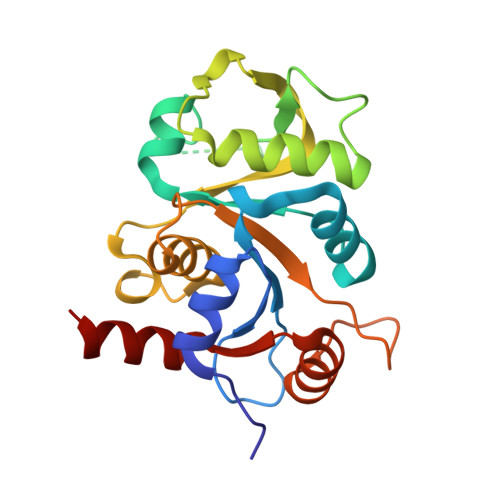
    3,4-dihydroxy 2-butanone 4-phosphate synthase (DHBPS) and GTP cyclohydrolase-II (GTPCH-II) are the two initial enzymes involved in riboflavin biosynthesis pathway, which has been shown to be essential for the pathogens. In Mycobacterium tuberculosis (Mtb), the ribA2 gene (Rv1415) encodes for the bi-functional enzyme with DHBPS and GTPCH-II domains at N- and C-termini, respectively. We have determined three crystal structures of Mtb-DHBPS domain in complex with phosphate and glycerol at pH 6.0, with sulphate at pH 4.0 and with zinc and sulphate at pH 4.0 at 1.8, 2.06 and 2.06 Å resolution, respectively. The hydrodynamic volume and enzyme activity studies revealed that the Mtb-DHBPS domain forms a functional homo-dimer between the pH 6.0 and 9.0, however, at pH 5.0 and below, it forms a stable inactive monomer in solution. Furthermore, the functional activity of Mtb-DHBPS and its dimeric state could be restored by increasing the pH between 6.0 and 9.0. The comparison of crystal structures determined at different pH revealed that the overall three-dimensional structure of Mtb-DHBPS monomer remains the same. However, the length of the α6-helix at pH 6.0 has increased from 15 to 22 Å in pH 4.0 by increasing the number of amino acids contributing to the α6-helix from 11 to 15, achieving a higher structural stability at pH 4.0. Taken together our experiments strongly suggest that the Mtb-DHBPS domain can transit between inactive monomer to active dimer depending upon its pH values, both in solution as well in crystal structure.


  • Organizational Affiliation

    Division of Protein Science and Engineering, Institute of Microbial Technology, Council of Scientific and Industrial Research (CSIR), Sector 39-A, Chandigarh 160036, India.


Macromolecules
Find similar proteins by:  (by identity cutoff)  |  3D Structure
Entity ID: 1
MoleculeChains Sequence LengthOrganismDetailsImage
3,4-dihydroxy-2-butanone 4-phosphate synthase
A, B
206Mycobacterium tuberculosis H37RaMutation(s): 0 
Gene Names: ribA2
EC: 4.1.99.12
UniProt
Find proteins for A5U2B7 (Mycobacterium tuberculosis (strain ATCC 25177 / H37Ra))
Explore A5U2B7 
Go to UniProtKB:  A5U2B7
Entity Groups  
Sequence Clusters30% Identity50% Identity70% Identity90% Identity95% Identity100% Identity
UniProt GroupA5U2B7
Sequence Annotations
Expand
  • Reference Sequence
Small Molecules
Ligands 3 Unique
IDChains Name / Formula / InChI Key2D Diagram3D Interactions
PO4
Query on PO4

Download Ideal Coordinates CCD File 
C [auth A],
D [auth A],
H [auth B]
PHOSPHATE ION
O4 P
NBIIXXVUZAFLBC-UHFFFAOYSA-K
GOL
Query on GOL

Download Ideal Coordinates CCD File 
E [auth A]
F [auth A]
I [auth B]
J [auth B]
K [auth B]
E [auth A],
F [auth A],
I [auth B],
J [auth B],
K [auth B],
L [auth B]
GLYCEROL
C3 H8 O3
PEDCQBHIVMGVHV-UHFFFAOYSA-N
K
Query on K

Download Ideal Coordinates CCD File 
G [auth A],
M [auth B]
POTASSIUM ION
K
NPYPAHLBTDXSSS-UHFFFAOYSA-N
Experimental Data & Validation

Experimental Data

  • Method: X-RAY DIFFRACTION
  • Resolution: 1.80 Å
  • R-Value Free: 0.233 
  • R-Value Work: 0.185 
  • R-Value Observed: 0.187 
  • Space Group: C 1 2 1
Unit Cell:
Length ( Å )Angle ( ˚ )
a = 89.784α = 90
b = 51.316β = 112.76
c = 95.407γ = 90
Software Package:
Software NamePurpose
MARMOSAICdata collection
PHASERphasing
REFMACrefinement
HKL-2000data reduction
HKL-2000data scaling

Structure Validation

View Full Validation Report


Entry History 

Deposition Data

Revision History  (Full details and data files)

  • Version 1.0: 2011-02-23
    Type: Initial release
  • Version 1.1: 2011-07-13
    Changes: Version format compliance
  • Version 1.2: 2011-12-07
    Changes: Database references
  • Version 1.3: 2023-11-01
    Changes: Data collection, Database references, Derived calculations, Refinement description