3M2K

Crystal Structure of fluorescein-labeled Class A -beta lactamase PenP in complex with cefotaxime


Experimental Data Snapshot

  • Method: X-RAY DIFFRACTION
  • Resolution: 3.50 Å
  • R-Value Free: 0.236 
  • R-Value Work: 0.176 
  • R-Value Observed: 0.180 

wwPDB Validation   3D Report Full Report


Ligand Structure Quality Assessment 


This is version 2.1 of the entry. See complete history


Literature

Structural studies of the mechanism for biosensing antibiotics in a fluorescein-labeled beta-lactamase.

Wong, W.T.Au, H.W.Yap, H.K.Leung, Y.C.Wong, K.Y.Zhao, Y.

(2011) BMC Struct Biol 11: 15-15

  • DOI: https://doi.org/10.1186/1472-6807-11-15
  • Primary Citation of Related Structures:  
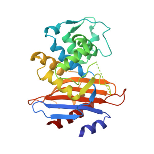
    3M2K

  • PubMed Abstract: 

    β-lactamase conjugated with environment-sensitive fluorescein molecule to residue 166 on the Ω-loop near its catalytic site is a highly effective biosensor for β-lactam antibiotics. Yet the molecular mechanism of such fluorescence-based biosensing is not well understood.


  • Organizational Affiliation

    Department of Applied Biology and Chemical Technology, Central Laboratory of Institute of Molecular Technology for Drug Discovery and Synthesis, The Hong Kong Polytechnic University, Hung Hom, Hong Hong, China.


Macromolecules
Find similar proteins by:  (by identity cutoff)  |  3D Structure
Entity ID: 1
MoleculeChains Sequence LengthOrganismDetailsImage
Beta-lactamase
A, B
257Bacillus licheniformisMutation(s): 1 
Gene Names: penP
EC: 3.5.2.6
UniProt
Find proteins for P00808 (Bacillus licheniformis)
Explore P00808 
Go to UniProtKB:  P00808
Entity Groups  
Sequence Clusters30% Identity50% Identity70% Identity90% Identity95% Identity100% Identity
UniProt GroupP00808
Sequence Annotations
Expand
  • Reference Sequence
Small Molecules
Ligands 1 Unique
IDChains Name / Formula / InChI Key2D Diagram3D Interactions
CEF
Query on CEF

Download Ideal Coordinates CCD File 
C [auth A],
D [auth B]
CEFOTAXIME, C3' cleaved, open, bound form
C14 H15 N5 O5 S2
NRYMPLKBKFIWQC-YVCCLBOHSA-N
Experimental Data & Validation

Experimental Data

  • Method: X-RAY DIFFRACTION
  • Resolution: 3.50 Å
  • R-Value Free: 0.236 
  • R-Value Work: 0.176 
  • R-Value Observed: 0.180 
  • Space Group: P 1 21 1
Unit Cell:
Length ( Å )Angle ( ˚ )
a = 43.538α = 90
b = 91.524β = 104.22
c = 66.302γ = 90
Software Package:
Software NamePurpose
CrystalCleardata collection
REFMACrefinement
CrystalCleardata reduction
CrystalCleardata scaling

Structure Validation

View Full Validation Report


Ligand Structure Quality Assessment 


Entry History 

Deposition Data

Revision History  (Full details and data files)

  • Version 1.0: 2011-03-16
    Type: Initial release
  • Version 1.1: 2011-07-13
    Changes: Version format compliance
  • Version 1.2: 2011-08-24
    Changes: Non-polymer description
  • Version 2.0: 2018-09-19
    Changes: Advisory, Atomic model, Data collection, Database references, Derived calculations, Non-polymer description, Structure summary
  • Version 2.1: 2023-11-01
    Changes: Data collection, Database references, Derived calculations, Refinement description