3LSP

Crystal Structure of DesT bound to desCB promoter and oleoyl-CoA


Experimental Data Snapshot

  • Method: X-RAY DIFFRACTION
  • Resolution: 2.66 Å
  • R-Value Free: 0.261 
  • R-Value Work: 0.223 
  • R-Value Observed: 0.225 

wwPDB Validation   3D Report Full Report


Ligand Structure Quality Assessment 


This is version 1.3 of the entry. See complete history


Literature

Structural basis for the transcriptional regulation of membrane lipid homeostasis.

Miller, D.J.Zhang, Y.M.Subramanian, C.Rock, C.O.White, S.W.

(2010) Nat Struct Mol Biol 17: 971-975

  • DOI: https://doi.org/10.1038/nsmb.1847
  • Primary Citation of Related Structures:  
    3LSJ, 3LSP, 3LSR

  • PubMed Abstract: 

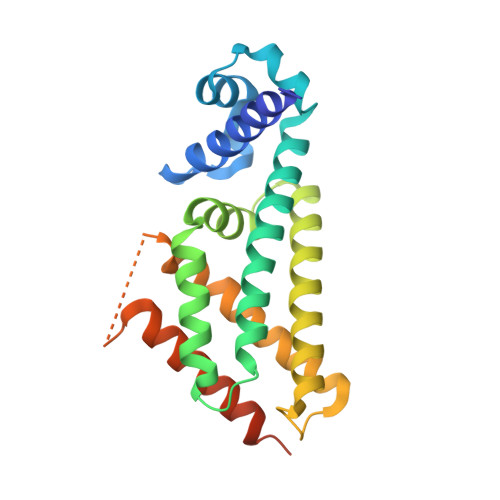
    DesT is a transcriptional repressor that regulates the genes that control the unsaturated:saturated fatty acid ratio available for membrane lipid synthesis. DesT bound to unsaturated acyl-CoA has a high affinity for its cognate palindromic DNA-binding site, whereas DesT bound to saturated acyl-CoA does not bind this site. Structural analyses of the DesT-oleoyl-CoA-DNA and DesT-palmitoyl-CoA complexes reveal that acyl chain shape directly influences the packing of hydrophobic core residues within the DesT ligand-binding domain. These changes are propagated to the paired DNA-binding domains via conformational changes to modulate DNA binding. These structural interpretations are supported by the in vitro and in vivo characterization of site-directed mutants. The regulation of DesT by the unsaturated:saturated ratio of acyl chains rather than the concentration of a single ligand is a paradigm for understanding transcriptional regulation of membrane lipid homeostasis.


  • Organizational Affiliation

    Department of Structural Biology, St. Jude Children's Research Hospital, Memphis, Tennessee, USA.


Macromolecules

Find similar proteins by:  (by identity cutoff)  |  3D Structure
Entity ID: 1
MoleculeChains Sequence LengthOrganismDetailsImage
DesT220Pseudomonas aeruginosaMutation(s): 1 
Gene Names: desTPA4890
UniProt
Find proteins for Q9HUS3 (Pseudomonas aeruginosa (strain ATCC 15692 / DSM 22644 / CIP 104116 / JCM 14847 / LMG 12228 / 1C / PRS 101 / PAO1))
Explore Q9HUS3 
Go to UniProtKB:  Q9HUS3
Entity Groups  
Sequence Clusters30% Identity50% Identity70% Identity90% Identity95% Identity100% Identity
UniProt GroupQ9HUS3
Sequence Annotations
Expand
  • Reference Sequence
Find similar nucleic acids by:  (by identity cutoff)  |  3D Structure
Entity ID: 2
MoleculeChains LengthOrganismImage
DNA (5'-D(*TP*TP*AP*CP*AP*TP*CP*AP*GP*TP*GP*AP*AP*CP*GP*CP*TP*TP*GP*TP*TP*GP*AP*CP*TP*CP*GP*AP*TP*TP*G)-3')31N/A
Sequence Annotations
Expand
  • Reference Sequence
Find similar nucleic acids by:  (by identity cutoff)  |  3D Structure
Entity ID: 3
MoleculeChains LengthOrganismImage
DNA (5'-D(*TP*CP*AP*AP*TP*CP*GP*AP*GP*TP*CP*AP*AP*CP*AP*AP*GP*CP*GP*TP*TP*CP*AP*CP*TP*GP*AP*TP*GP*TP*A)-3')C [auth D]31N/A
Sequence Annotations
Expand
  • Reference Sequence
Experimental Data & Validation

Experimental Data

  • Method: X-RAY DIFFRACTION
  • Resolution: 2.66 Å
  • R-Value Free: 0.261 
  • R-Value Work: 0.223 
  • R-Value Observed: 0.225 
  • Space Group: I 41
Unit Cell:
Length ( Å )Angle ( ˚ )
a = 78.385α = 90
b = 78.385β = 90
c = 147.988γ = 90
Software Package:
Software NamePurpose
HKL-2000data collection
PHASERphasing
REFMACrefinement
HKL-2000data reduction
HKL-2000data scaling

Structure Validation

View Full Validation Report


Ligand Structure Quality Assessment 


Entry History 

Deposition Data

Revision History  (Full details and data files)

  • Version 1.0: 2010-08-04
    Type: Initial release
  • Version 1.1: 2011-07-13
    Changes: Version format compliance
  • Version 1.2: 2021-10-13
    Changes: Database references, Derived calculations
  • Version 1.3: 2024-02-21
    Changes: Data collection