


1.8 Angstrom Resolution Crystal Structure of Enoyl-CoA Hydratase from Bacillus anthracis.
Minasov, G., Halavaty, A., Wawrzak, Z., Skarina, T., Onopriyenko, O., Papazisi, L., Savchenko, A., Anderson, W.F.To be published.
Experimental Data Snapshot
wwPDB Validation 3D Report Full Report

Entity ID: 1 | |||||
|---|---|---|---|---|---|
| Molecule | Chains | Sequence Length | Organism | Details | Image |
| Enoyl-CoA hydratase/isomerase family protein | 265 | Bacillus anthracis | Mutation(s): 0 Gene Names: BAS2374, BA_2551, GBAA2551, GBAA_2551 EC: 4.2.1.17 | 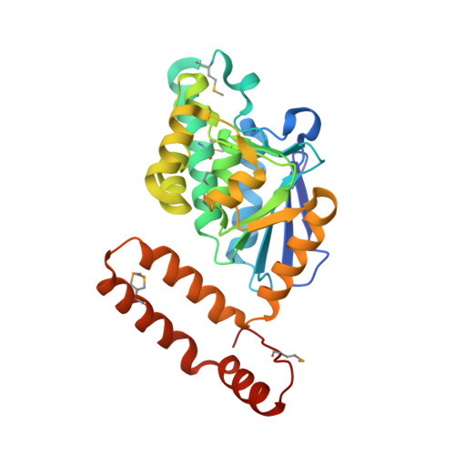 | |
UniProt | |||||
Find proteins for A0A6L8PDF4 (Bacillus anthracis) Explore A0A6L8PDF4 Go to UniProtKB: A0A6L8PDF4 | |||||
Entity Groups | |||||
| Sequence Clusters | 30% Identity50% Identity70% Identity90% Identity95% Identity100% Identity | ||||
| UniProt Group | A0A6L8PDF4 | ||||
Sequence AnnotationsExpand | |||||
| |||||
| Ligands 2 Unique | |||||
|---|---|---|---|---|---|
| ID | Chains | Name / Formula / InChI Key | 2D Diagram | 3D Interactions | |
| CA Query on CA | G [auth A], H [auth D], I [auth E], L [auth F], M [auth F] | CALCIUM ION Ca BHPQYMZQTOCNFJ-UHFFFAOYSA-N |  | ||
| CL Query on CL | J [auth E], K [auth E] | CHLORIDE ION Cl VEXZGXHMUGYJMC-UHFFFAOYSA-M |  | ||
| Modified Residues 1 Unique | |||||
|---|---|---|---|---|---|
| ID | Chains | Type | Formula | 2D Diagram | Parent |
| MSE Query on MSE | A, B, C, D, E A, B, C, D, E, F | L-PEPTIDE LINKING | C5 H11 N O2 Se |  | MET |
| Length ( Å ) | Angle ( ˚ ) |
|---|---|
| a = 73.747 | α = 90 |
| b = 130.721 | β = 114.47 |
| c = 73.802 | γ = 90 |
| Software Name | Purpose |
|---|---|
| Blu-Ice | data collection |
| CRANK | phasing |
| REFMAC | refinement |
| HKL-2000 | data reduction |
| HKL-2000 | data scaling |
RCSB PDB (citation) is hosted by
RCSB PDB is a member of the

