Crystal structure of the cofactor-independent monooxygenase SnoaB from Streptomyces nogalater: implications for the reaction mechanism
Grocholski, T., Koskiniemi, H., Lindqvist, Y., Mantsala, P., Niemi, J., Schneider, G.(2010) Biochemistry 49: 934-944
- PubMed: 20052967
- DOI: https://doi.org/10.1021/bi901985b
- Primary Citation of Related Structures:
3KG0, 3KG1, 3KNG - PubMed Abstract:
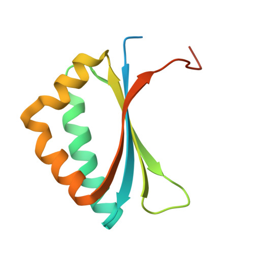
SnoaB is a cofactor-independent monooxygenase that catalyzes the conversion of 12-deoxynogalonic acid to nogalonic acid in the biosynthesis of the aromatic polyketide nogalamycin in Streptomyces nogalater. In vitro (18)O(2) experiments establish that the oxygen atom incorporated into the substrate is derived from molecular oxygen. The crystal structure of the enzyme was determined in two different space groups to 1.7 and 1.9 A resolution, respectively. The enzyme displays the ferredoxin fold, with the characteristic beta-strand exchange at the dimer interface. The crystal structures reveal a putative catalytic triad involving two asparagine residues, Asn18 and Asn63, and a water molecule, which may play important roles in the enzymatic reaction. Site-directed mutagenesis experiments, replacing the two asparagines individually by alanine, led to a 100-fold drop in enzymatic activity. Replacement of an invariant tryptophan residue in the active site of the enzyme by phenylalanine also resulted in an enzyme variant with about 1% residual activity. Taken together, our findings are most consistent with a carbanion mechanism where the deprotonated substrate reacts with molecular oxygen via one electron transfer and formation of a caged radical.
Organizational Affiliation:
Department of Biochemistry and Food Chemistry, University of Turku, FIN-20014 Turku, Finland.