3K9P

The crystal structure of E2-25K and ubiquitin complex


Experimental Data Snapshot

  • Method: X-RAY DIFFRACTION
  • Resolution: 2.80 Å
  • R-Value Free: 0.296 
  • R-Value Work: 0.232 
  • R-Value Observed: 0.232 

wwPDB Validation   3D Report Full Report


This is version 1.2 of the entry. See complete history


Literature

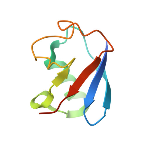
Structural basis of E2-25K/UBB+1 interaction leading to proteasome inhibition and neurotoxicity

Ko, S.Kang, G.B.Song, S.M.Lee, J.-G.Shin, D.Y.Yun, J.-H.Sheng, Y.Cheong, C.Jeon, Y.H.Jung, Y.-K.Arrowsmith, C.H.Avvakumov, G.V.Dhe-Paganon, S.Yoo, Y.J.Eom, S.H.Lee, W.

(2010) J Biol Chem 285: 36070-36080

  • DOI: https://doi.org/10.1074/jbc.M110.145219
  • Primary Citation of Related Structures:  
    1YLA, 3K9P

  • PubMed Abstract: 

    E2-25K/Hip2 is an unusual ubiquitin-conjugating enzyme that interacts with the frameshift mutant of ubiquitin B (UBB(+1)) and has been identified as a crucial factor regulating amyloid-β neurotoxicity. To study the structural basis of the neurotoxicity mediated by the E2-25K-UBB(+1) interaction, we determined the three-dimensional structures of UBB(+1), E2-25K and the E2-25K/ubiquitin, and E2-25K/UBB(+1) complex. The structures revealed that ubiquitin or UBB(+1) is bound to E2-25K via the enzyme MGF motif and residues in α9 of the enzyme. Polyubiquitylation assays together with analyses of various E2-25K mutants showed that disrupting UBB(+1) binding markedly diminishes synthesis of neurotoxic UBB(+1)-anchored polyubiquitin. These results suggest that the interaction between E2-25K and UBB(+1) is critical for the synthesis and accumulation of UBB(+1)-anchored polyubiquitin, which results in proteasomal inhibition and neuronal cell death.


  • Organizational Affiliation

    Department of Biochemistry, College of Life Science and Biotechnology, Yonsei University, Seoul 120-749, Korea.


Macromolecules
Find similar proteins by:  (by identity cutoff)  |  3D Structure
Entity ID: 1
MoleculeChains Sequence LengthOrganismDetailsImage
Ubiquitin-conjugating enzyme E2 K217Homo sapiensMutation(s): 0 
Gene Names: UBE2KHIP2LIG
EC: 6.3.2.19
UniProt & NIH Common Fund Data Resources
Find proteins for P61086 (Homo sapiens)
Explore P61086 
Go to UniProtKB:  P61086
PHAROS:  P61086
GTEx:  ENSG00000078140 
Entity Groups  
Sequence Clusters30% Identity50% Identity70% Identity90% Identity95% Identity100% Identity
UniProt GroupP61086
Sequence Annotations
Expand
  • Reference Sequence
Find similar proteins by:  (by identity cutoff)  |  3D Structure
Entity ID: 2
MoleculeChains Sequence LengthOrganismDetailsImage
Ubiquitin79Homo sapiensMutation(s): 0 
UniProt & NIH Common Fund Data Resources
Find proteins for P0CG48 (Homo sapiens)
Explore P0CG48 
Go to UniProtKB:  P0CG48
PHAROS:  P0CG48
GTEx:  ENSG00000150991 
Entity Groups  
Sequence Clusters30% Identity50% Identity70% Identity90% Identity95% Identity100% Identity
UniProt GroupP0CG48
Sequence Annotations
Expand
  • Reference Sequence
Experimental Data & Validation

Experimental Data

  • Method: X-RAY DIFFRACTION
  • Resolution: 2.80 Å
  • R-Value Free: 0.296 
  • R-Value Work: 0.232 
  • R-Value Observed: 0.232 
  • Space Group: P 1 21 1
Unit Cell:
Length ( Å )Angle ( ˚ )
a = 43.554α = 90
b = 50.974β = 102.62
c = 63.906γ = 90
Software Package:
Software NamePurpose
HKL-2000data collection
MOLREPphasing
CNSrefinement
HKL-2000data reduction
HKL-2000data scaling

Structure Validation

View Full Validation Report


Entry History 

Deposition Data

Revision History  (Full details and data files)

  • Version 1.0: 2010-09-08
    Type: Initial release
  • Version 1.1: 2011-07-13
    Changes: Version format compliance
  • Version 1.2: 2024-03-20
    Changes: Data collection, Database references