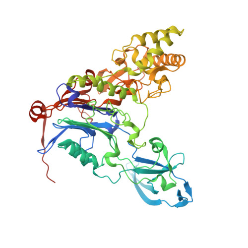
3JTR

Mutations in Cephalosporin Acylase Affecting Stability and Autoproteolysis


Experimental Data Snapshot

  • Method: X-RAY DIFFRACTION
  • Resolution: 2.50 Å
  • R-Value Free: 0.224 
  • R-Value Work: 0.182 
  • R-Value Observed: 0.184 

wwPDB Validation   3D Report Full Report


This is version 1.4 of the entry. See complete history


Literature

Structural features of cephalosporin acylase reveal the basis of autocatalytic activation.

Cho, K.J.Kim, J.K.Lee, J.H.Shin, H.J.Park, S.S.Kim, K.H.

(2009) Biochem Biophys Res Commun 390: 342-348

  • DOI: https://doi.org/10.1016/j.bbrc.2009.09.134
  • Primary Citation of Related Structures:  
    3JTQ, 3JTR

  • PubMed Abstract: 

    Cephalosporin acylase (CA), a member of the N-terminal nucleophile hydrolase family, is activated through two steps of intramolecular autoproteolysis, the first mediated by a serine residue, and the second by a glutamate, which releases the pro-segment and produces an active enzyme. In this study, we have determined the crystal structures of mutants which could affect primary or secondary auto-cleavage and of sequential intermediates of a slow-processing mutant at 2.0-2.5A resolutions. The pro-segments of the mutants undergo dynamic conformational changes during activation and adopt surprisingly different loop conformations from one another. However, the autoproteolytic site was found to form a catalytically competent conformation with a solvent water molecule, which was essentially conserved in the CA mutants.


  • Organizational Affiliation

    Department of Life Sciences and Biotechnology, Korea University, Seoul 136-701, Republic of Korea.


Macromolecules
Find similar proteins by:  (by identity cutoff)  |  3D Structure
Entity ID: 1
MoleculeChains Sequence LengthOrganismDetailsImage
Glutaryl 7-aminocephalosporanic acid acylase169Pseudomonas sp. GK16Mutation(s): 1 
EC: 3.5.1.93
UniProt
Find proteins for A4ZVL3 (Pseudomonas sp. GK16)
Explore A4ZVL3 
Go to UniProtKB:  A4ZVL3
Entity Groups  
Sequence Clusters30% Identity50% Identity70% Identity90% Identity95% Identity100% Identity
UniProt GroupA4ZVL3
Sequence Annotations
Expand
  • Reference Sequence
Find similar proteins by:  (by identity cutoff)  |  3D Structure
Entity ID: 2
MoleculeChains Sequence LengthOrganismDetailsImage
Glutaryl 7-aminocephalosporanic acid acylase528Pseudomonas sp. GK16Mutation(s): 0 
EC: 3.5.1.93
UniProt
Find proteins for A4ZVL3 (Pseudomonas sp. GK16)
Explore A4ZVL3 
Go to UniProtKB:  A4ZVL3
Entity Groups  
Sequence Clusters30% Identity50% Identity70% Identity90% Identity95% Identity100% Identity
UniProt GroupA4ZVL3
Sequence Annotations
Expand
  • Reference Sequence
Small Molecules
Ligands 1 Unique
IDChains Name / Formula / InChI Key2D Diagram3D Interactions
GOL
Query on GOL

Download Ideal Coordinates CCD File 
C [auth B]GLYCEROL
C3 H8 O3
PEDCQBHIVMGVHV-UHFFFAOYSA-N
Experimental Data & Validation

Experimental Data

  • Method: X-RAY DIFFRACTION
  • Resolution: 2.50 Å
  • R-Value Free: 0.224 
  • R-Value Work: 0.182 
  • R-Value Observed: 0.184 
  • Space Group: P 41 21 2
Unit Cell:
Length ( Å )Angle ( ˚ )
a = 73.816α = 90
b = 73.816β = 90
c = 381.324γ = 90
Software Package:
Software NamePurpose
DENZOdata reduction
SCALEPACKdata scaling
REFMACrefinement
PDB_EXTRACTdata extraction
HKL-2000data collection
HKL-2000data reduction
HKL-2000data scaling
CNSphasing

Structure Validation

View Full Validation Report


Entry History 

Deposition Data

Revision History  (Full details and data files)

  • Version 1.0: 2010-01-26
    Type: Initial release
  • Version 1.1: 2011-07-13
    Changes: Version format compliance
  • Version 1.2: 2017-11-01
    Changes: Refinement description
  • Version 1.3: 2021-11-10
    Changes: Database references, Derived calculations
  • Version 1.4: 2023-11-01
    Changes: Data collection, Refinement description