3IQD

Structure of Octopine-dehydrogenase in complex with NADH and Agmatine


Experimental Data Snapshot

  • Method: X-RAY DIFFRACTION
  • Resolution: 2.80 Å
  • R-Value Free: 0.257 
  • R-Value Work: 0.202 
  • R-Value Observed: 0.205 

wwPDB Validation   3D Report Full Report


Ligand Structure Quality Assessment 


This is version 1.3 of the entry. See complete history


Literature

Insights into the mechanism of ligand binding to octopine dehydrogenase from Pecten maximus by NMR and crystallography

Smits, S.H.J.Meyer, T.Mueller, A.van Os, N.Stoldt, M.Willbold, D.Schmitt, L.Grieshaber, M.K.

(2010) PLoS One 5: e12312-e12312

  • DOI: https://doi.org/10.1371/journal.pone.0012312
  • Primary Citation of Related Structures:  
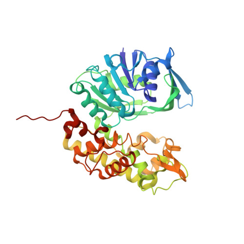
    3IQD

  • PubMed Abstract: 

    Octopine dehydrogenase (OcDH) from the adductor muscle of the great scallop, Pecten maximus, catalyzes the NADH dependent, reductive condensation of L-arginine and pyruvate to octopine, NAD(+), and water during escape swimming and/or subsequent recovery. The structure of OcDH was recently solved and a reaction mechanism was proposed which implied an ordered binding of NADH, L-arginine and finally pyruvate. Here, the order of substrate binding as well as the underlying conformational changes were investigated by NMR confirming the model derived from the crystal structures. Furthermore, the crystal structure of the OcDH/NADH/agmatine complex was determined which suggests a key role of the side chain of L-arginine in protein cataylsis. Thus, the order of substrate binding to OcDH as well as the molecular signals involved in octopine formation can now be described in molecular detail.


  • Organizational Affiliation

    Institute of Biochemistry, Heinrich-Heine-University, Duesseldorf, Germany. sander.smits@uni-duesseldorf.de


Macromolecules
Find similar proteins by:  (by identity cutoff)  |  3D Structure
Entity ID: 1
MoleculeChains Sequence LengthOrganismDetailsImage
Octopine dehydrogenaseA [auth B]404Pecten maximusMutation(s): 0 
Gene Names: odh1
EC: 1.5.1.11
UniProt
Find proteins for Q9BHM6 (Pecten maximus)
Explore Q9BHM6 
Go to UniProtKB:  Q9BHM6
Entity Groups  
Sequence Clusters30% Identity50% Identity70% Identity90% Identity95% Identity100% Identity
UniProt GroupQ9BHM6
Sequence Annotations
Expand
  • Reference Sequence
Small Molecules
Ligands 2 Unique
IDChains Name / Formula / InChI Key2D Diagram3D Interactions
NAI
Query on NAI

Download Ideal Coordinates CCD File 
B
1,4-DIHYDRONICOTINAMIDE ADENINE DINUCLEOTIDE
C21 H29 N7 O14 P2
BOPGDPNILDQYTO-NNYOXOHSSA-N
AG2
Query on AG2

Download Ideal Coordinates CCD File 
C [auth B]AGMATINE
C5 H14 N4
QYPPJABKJHAVHS-UHFFFAOYSA-N
Experimental Data & Validation

Experimental Data

  • Method: X-RAY DIFFRACTION
  • Resolution: 2.80 Å
  • R-Value Free: 0.257 
  • R-Value Work: 0.202 
  • R-Value Observed: 0.205 
  • Space Group: P 41 21 2
Unit Cell:
Length ( Å )Angle ( ˚ )
a = 96.2α = 90
b = 96.2β = 90
c = 117.4γ = 90
Software Package:
Software NamePurpose
DNAdata collection
PHASERphasing
REFMACrefinement
XDSdata reduction
XSCALEdata scaling

Structure Validation

View Full Validation Report


Ligand Structure Quality Assessment 


Entry History 

Deposition Data

Revision History  (Full details and data files)

  • Version 1.0: 2010-08-25
    Type: Initial release
  • Version 1.1: 2011-07-13
    Changes: Version format compliance
  • Version 1.2: 2013-11-06
    Changes: Non-polymer description
  • Version 1.3: 2023-11-01
    Changes: Data collection, Database references, Derived calculations, Refinement description