3IKM

Crystal structure of human mitochondrial DNA polymerase holoenzyme


Experimental Data Snapshot

  • Method: X-RAY DIFFRACTION
  • Resolution: 3.24 Å
  • R-Value Free: 0.303 
  • R-Value Work: 0.284 
  • R-Value Observed: 0.284 

wwPDB Validation   3D Report Full Report


This is version 1.3 of the entry. See complete history


Literature

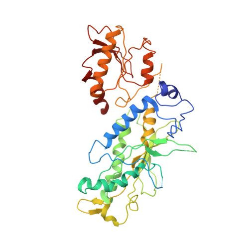
Structural insight into processive human mitochondrial DNA synthesis and disease-related polymerase mutations.

Lee, Y.S.Kennedy, W.D.Yin, Y.W.

(2009) Cell 139: 312-324

  • DOI: https://doi.org/10.1016/j.cell.2009.07.050
  • Primary Citation of Related Structures:  
    3IKL, 3IKM

  • PubMed Abstract: 

    Human mitochondrial DNA polymerase (Pol gamma) is the sole replicase in mitochondria. Pol gamma is vulnerable to nonselective antiretroviral drugs and is increasingly associated with mutations found in patients with mitochondriopathies. We determined crystal structures of the human heterotrimeric Pol gamma holoenzyme and, separately, a variant of its processivity factor, Pol gammaB. The holoenzyme structure reveals an unexpected assembly of the mitochondrial DNA replicase where the catalytic subunit Pol gammaA interacts with its processivity factor primarily via a domain that is absent in all other DNA polymerases. This domain provides a structural module for supporting both the intrinsic processivity of the catalytic subunit alone and the enhanced processivity of holoenzyme. The Pol gamma structure also provides a context for interpreting the phenotypes of disease-related mutations in the polymerase and establishes a foundation for understanding the molecular basis of toxicity of anti-retroviral drugs targeting HIV reverse transcriptase.


  • Organizational Affiliation

    Institute for Cellular and Molecular Biology, University of Texas at Austin, Austin, TX 78712, USA.


Macromolecules
Find similar proteins by:  (by identity cutoff)  |  3D Structure
Entity ID: 1
MoleculeChains Sequence LengthOrganismDetailsImage
DNA polymerase subunit gamma-1
A, D
1,172Homo sapiensMutation(s): 0 
EC: 2.7.7.7
UniProt & NIH Common Fund Data Resources
Find proteins for P54098 (Homo sapiens)
Explore P54098 
Go to UniProtKB:  P54098
PHAROS:  P54098
GTEx:  ENSG00000140521 
Entity Groups  
Sequence Clusters30% Identity50% Identity70% Identity90% Identity95% Identity100% Identity
UniProt GroupP54098
Sequence Annotations
Expand
  • Reference Sequence
Find similar proteins by:  (by identity cutoff)  |  3D Structure
Entity ID: 2
MoleculeChains Sequence LengthOrganismDetailsImage
DNA polymerase subunit gamma-2
B, C, E, F
427Homo sapiensMutation(s): 0 
EC: 2.7.7.7
UniProt & NIH Common Fund Data Resources
Find proteins for Q9UHN1 (Homo sapiens)
Explore Q9UHN1 
Go to UniProtKB:  Q9UHN1
PHAROS:  Q9UHN1
GTEx:  ENSG00000256525 
Entity Groups  
Sequence Clusters30% Identity50% Identity70% Identity90% Identity95% Identity100% Identity
UniProt GroupQ9UHN1
Sequence Annotations
Expand
  • Reference Sequence
Experimental Data & Validation

Experimental Data

  • Method: X-RAY DIFFRACTION
  • Resolution: 3.24 Å
  • R-Value Free: 0.303 
  • R-Value Work: 0.284 
  • R-Value Observed: 0.284 
  • Space Group: P 32
Unit Cell:
Length ( Å )Angle ( ˚ )
a = 138.394α = 90
b = 138.394β = 90
c = 226.312γ = 120
Software Package:
Software NamePurpose
ADSCdata collection
SOLVEphasing
CNSrefinement
HKL-2000data reduction
HKL-2000data scaling

Structure Validation

View Full Validation Report


Entry History 

Deposition Data

Revision History  (Full details and data files)

  • Version 1.0: 2009-11-03
    Type: Initial release
  • Version 1.1: 2011-07-13
    Changes: Version format compliance
  • Version 1.2: 2018-01-24
    Changes: Structure summary
  • Version 1.3: 2024-02-21
    Changes: Data collection, Database references