3I00

Crystal Structure of the huntingtin interacting protein 1 coiled coil domain


Experimental Data Snapshot

  • Method: X-RAY DIFFRACTION
  • Resolution: 2.30 Å
  • R-Value Free: 0.280 
  • R-Value Work: 0.235 
  • R-Value Observed: 0.237 

wwPDB Validation   3D Report Full Report


This is version 1.3 of the entry. See complete history


Literature

Accommodation of structural rearrangements in the huntingtin-interacting protein 1 coiled-coil domain.

Wilbur, J.D.Hwang, P.K.Brodsky, F.M.Fletterick, R.J.

(2010) Acta Crystallogr D Biol Crystallogr 66: 314-318

  • DOI: https://doi.org/10.1107/S0907444909054535
  • Primary Citation of Related Structures:  
    3I00

  • PubMed Abstract: 

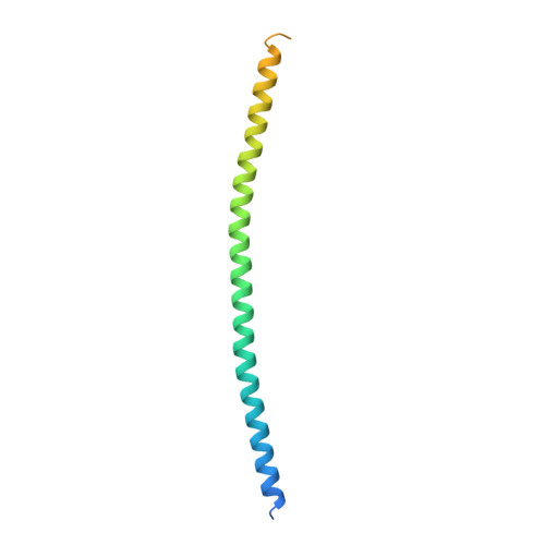
    Huntingtin-interacting protein 1 (HIP1) is an important link between the actin cytoskeleton and clathrin-mediated endocytosis machinery. HIP1 has also been implicated in the pathogenesis of Huntington's disease. The binding of HIP1 to actin is regulated through an interaction with clathrin light chain. Clathrin light chain binds to a flexible coiled-coil domain in HIP1 and induces a compact state that is refractory to actin binding. To understand the mechanism of this conformational regulation, a high-resolution crystal structure of a stable fragment from the HIP1 coiled-coil domain was determined. The flexibility of the HIP1 coiled-coil region was evident from its variation from a previously determined structure of a similar region. A hydrogen-bond network and changes in coiled-coil monomer interaction suggest that the HIP1 coiled-coil domain is uniquely suited to allow conformational flexibility.


  • Organizational Affiliation

    Graduate Program in Biophysics, University of California, San Francisco, California 94143, USA. jwilbur@msg.ucsf.edu


Macromolecules
Find similar proteins by:  (by identity cutoff)  |  3D Structure
Entity ID: 1
MoleculeChains Sequence LengthOrganismDetailsImage
Huntingtin-interacting protein 1
A, B
120Homo sapiensMutation(s): 0 
Gene Names: HIP1huntingtin interactin protein 1 (HIP1)
UniProt & NIH Common Fund Data Resources
Find proteins for O00291 (Homo sapiens)
Explore O00291 
Go to UniProtKB:  O00291
PHAROS:  O00291
GTEx:  ENSG00000127946 
Entity Groups  
Sequence Clusters30% Identity50% Identity70% Identity90% Identity95% Identity100% Identity
UniProt GroupO00291
Sequence Annotations
Expand
  • Reference Sequence
Experimental Data & Validation

Experimental Data

  • Method: X-RAY DIFFRACTION
  • Resolution: 2.30 Å
  • R-Value Free: 0.280 
  • R-Value Work: 0.235 
  • R-Value Observed: 0.237 
  • Space Group: P 21 21 21
Unit Cell:
Length ( Å )Angle ( ˚ )
a = 35.801α = 90
b = 57.092β = 90
c = 81.161γ = 90
Software Package:
Software NamePurpose
DENZOdata reduction
SCALEPACKdata scaling
PHASERphasing
REFMACrefinement
PDB_EXTRACTdata extraction
HKL-2000data reduction
HKL-2000data scaling

Structure Validation

View Full Validation Report


Entry History 

Deposition Data

Revision History  (Full details and data files)

  • Version 1.0: 2010-02-23
    Type: Initial release
  • Version 1.1: 2011-07-13
    Changes: Version format compliance
  • Version 1.2: 2012-03-21
    Changes: Database references
  • Version 1.3: 2017-11-01
    Changes: Refinement description