3HZA

Crystal structure of dUTPase H145W mutant


Experimental Data Snapshot

  • Method: X-RAY DIFFRACTION
  • Resolution: 1.20 Å
  • R-Value Free: 0.159 
  • R-Value Work: 0.117 
  • R-Value Observed: 0.117 

wwPDB Validation   3D Report Full Report


Ligand Structure Quality Assessment 


This is version 1.4 of the entry. See complete history


Literature

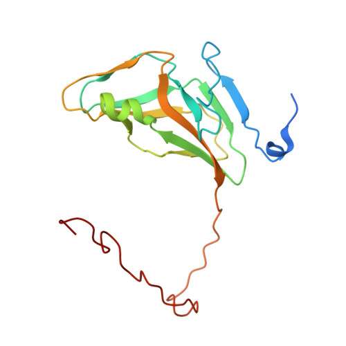
Aromatic stacking between nucleobase and enzyme promotes phosphate ester hydrolysis in dUTPase.

Pecsi, I.Leveles, I.Harmat, V.Vertessy, B.G.Toth, J.

(2010) Nucleic Acids Res 38: 7179-7186

  • DOI: https://doi.org/10.1093/nar/gkq584
  • Primary Citation of Related Structures:  
    3HZA, 3LOJ

  • PubMed Abstract: 

    Aromatic interactions are well-known players in molecular recognition but their catalytic role in biological systems is less documented. Here, we report that a conserved aromatic stacking interaction between dUTPase and its nucleotide substrate largely contributes to the stabilization of the associative type transition state of the nucleotide hydrolysis reaction. The effect of the aromatic stacking on catalysis is peculiar in that uracil, the aromatic moiety influenced by the aromatic interaction is relatively distant from the site of hydrolysis at the alpha-phosphate group. Using crystallographic, kinetics, optical spectroscopy and thermodynamics calculation approaches we delineate a possible mechanism by which rate acceleration is achieved through the remote π-π interaction. The abundance of similarly positioned aromatic interactions in various nucleotide hydrolyzing enzymes (e.g. most families of ATPases) raises the possibility of the reported phenomenon being a general component of the enzymatic catalysis of phosphate ester hydrolysis.


  • Organizational Affiliation

    Institute of Enzymology, Biological Research Center, Hungarian Academy of Sciences, Budapest, Hungary.


Macromolecules
Find similar proteins by:  (by identity cutoff)  |  3D Structure
Entity ID: 1
MoleculeChains Sequence LengthOrganismDetailsImage
Deoxyuridine 5'-triphosphate nucleotidohydrolase174Mycobacterium tuberculosisMutation(s): 1 
Gene Names: dutRv2697cMT2771MTCY05A6.18c
EC: 3.6.1.23
UniProt
Find proteins for P9WNS5 (Mycobacterium tuberculosis (strain ATCC 25618 / H37Rv))
Explore P9WNS5 
Go to UniProtKB:  P9WNS5
Entity Groups  
Sequence Clusters30% Identity50% Identity70% Identity90% Identity95% Identity100% Identity
UniProt GroupP9WNS5
Sequence Annotations
Expand
  • Reference Sequence
Experimental Data & Validation

Experimental Data

  • Method: X-RAY DIFFRACTION
  • Resolution: 1.20 Å
  • R-Value Free: 0.159 
  • R-Value Work: 0.117 
  • R-Value Observed: 0.117 
  • Space Group: P 63
Unit Cell:
Length ( Å )Angle ( ˚ )
a = 55.058α = 90
b = 55.058β = 90
c = 83.793γ = 120
Software Package:
Software NamePurpose
MAR345data collection
MOLREPphasing
SHELXL-97refinement
XDSdata reduction
XSCALEdata scaling

Structure Validation

View Full Validation Report


Ligand Structure Quality Assessment 


Entry History 

Deposition Data

Revision History  (Full details and data files)

  • Version 1.0: 2009-11-24
    Type: Initial release
  • Version 1.1: 2011-07-13
    Changes: Non-polymer description, Version format compliance
  • Version 1.2: 2017-11-01
    Changes: Advisory, Refinement description
  • Version 1.3: 2021-10-13
    Changes: Advisory, Data collection, Database references, Derived calculations
  • Version 1.4: 2023-09-06
    Changes: Data collection, Refinement description