3HUD

THE STRUCTURE OF HUMAN BETA 1 BETA 1 ALCOHOL DEHYDROGENASE: CATALYTIC EFFECTS OF NON-ACTIVE-SITE SUBSTITUTIONS


Experimental Data Snapshot

  • Method: X-RAY DIFFRACTION
  • Resolution: 3.20 Å
  • R-Value Work: 0.179 
  • R-Value Observed: 0.179 

wwPDB Validation   3D Report Full Report


This is version 1.4 of the entry. See complete history


Literature

Structure of human beta 1 beta 1 alcohol dehydrogenase: catalytic effects of non-active-site substitutions.

Hurley, T.D.Bosron, W.F.Hamilton, J.A.Amzel, L.M.

(1991) Proc Natl Acad Sci U S A 88: 8149-8153

  • DOI: https://doi.org/10.1073/pnas.88.18.8149
  • Primary Citation of Related Structures:  
    3HUD

  • PubMed Abstract: 

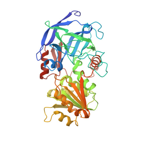
    The three-dimensional structure of human beta 1 beta 1 alcohol dehydrogenase (ADH; EC 1.1.1.1) complexed with NAD+ has been determined by x-ray crystallography to 3.0-A resolution. The amino acids directly involved in coenzyme binding are conserved between horse EE and human beta 1 beta 1 alcohol dehydrogenase in all but one case [serine (horse) vs. threonine (human) at position 48]. As a result, the coenzyme molecule is bound in a similar manner in the two enzymes. However, the strength of the interactions in the vicinity of the pyrophosphate bridge of NAD+ appears to be enhanced in the human enzyme. Side-chain movements of Arg-47 and Asp-50 and a shift in the position of the helix comprising residues 202-212 may explain both the decreased Vmax and the decreased rate of NADH dissociation observed in the human enzyme vs. the horse enzyme. It appears that these catalytic differences are not due to substitutions of any amino acids directly involved in coenzyme binding but are the result of structural rearrangements resulting from multiple sequence differences between the two enzymes.


  • Organizational Affiliation

    Department of Biochemistry and Molecular Biology, Indiana University School of Medicine, Indianapolis 46202.


Macromolecules
Find similar proteins by:  (by identity cutoff)  |  3D Structure
Entity ID: 1
MoleculeChains Sequence LengthOrganismDetailsImage
ALCOHOL DEHYDROGENASE
A, B
374Homo sapiensMutation(s): 0 
EC: 1.1.1.1
UniProt & NIH Common Fund Data Resources
Find proteins for P00325 (Homo sapiens)
Explore P00325 
Go to UniProtKB:  P00325
PHAROS:  P00325
GTEx:  ENSG00000196616 
Entity Groups  
Sequence Clusters30% Identity50% Identity70% Identity90% Identity95% Identity100% Identity
UniProt GroupP00325
Sequence Annotations
Expand
  • Reference Sequence
Experimental Data & Validation

Experimental Data

  • Method: X-RAY DIFFRACTION
  • Resolution: 3.20 Å
  • R-Value Work: 0.179 
  • R-Value Observed: 0.179 
  • Space Group: P 1
Unit Cell:
Length ( Å )Angle ( ˚ )
a = 54.59α = 91.93
b = 45.05β = 102.5
c = 93.86γ = 68.82
Software Package:
Software NamePurpose
X-PLORmodel building
X-PLORrefinement
X-PLORphasing

Structure Validation

View Full Validation Report


Entry History 

Deposition Data

Revision History  (Full details and data files)

  • Version 1.0: 1994-01-31
    Type: Initial release
  • Version 1.1: 2008-03-03
    Changes: Version format compliance
  • Version 1.2: 2011-07-13
    Changes: Version format compliance
  • Version 1.3: 2017-11-29
    Changes: Derived calculations, Other
  • Version 1.4: 2024-02-21
    Changes: Data collection, Database references, Derived calculations