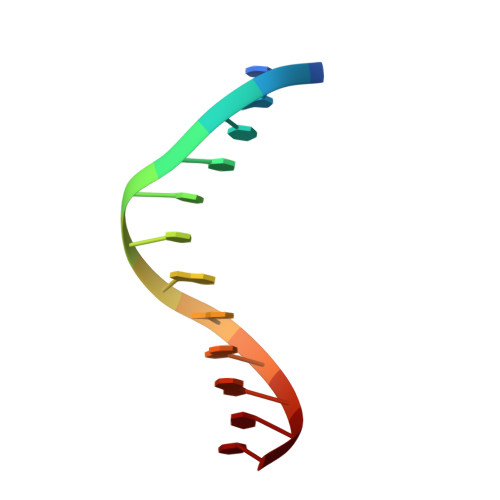
3HTS

HEAT SHOCK TRANSCRIPTION FACTOR/DNA COMPLEX


Experimental Data Snapshot

  • Method: X-RAY DIFFRACTION
  • Resolution: 1.75 Å
  • R-Value Free: 0.245 
  • R-Value Work: 0.208 

wwPDB Validation   3D Report Full Report


This is version 1.4 of the entry. See complete history


Literature

A new use for the 'wing' of the 'winged' helix-turn-helix motif in the HSF-DNA cocrystal.

Littlefield, O.Nelson, H.C.

(1999) Nat Struct Biol 6: 464-470

  • DOI: https://doi.org/10.1038/8269
  • Primary Citation of Related Structures:  
    3HTS

  • PubMed Abstract: 

    The 1.75 A crystal structure of the Kluyveromyces lactis heat shock transcription factor (HSF) DNA-binding domain (DBD) complexed with DNA reveals a protein-DNA interface with few direct major groove contacts and a number of phosphate backbone contacts that are primarily water-mediated interactions. The DBD, a 'winged' helix-turn-helix protein, displays a novel mode of binding in that the 'wing' does not contact DNA like all others of that class. Instead, the monomeric DBD, which crystallized as a symmetric dimer to a pair of nGAAn inverted repeats, uses the 'wing' to form part of the protein-protein contacts. This dimer interface is likely important for increasing the DNA-binding specificity and affinity of the trimeric form of HSF, as well as for increasing cooperativity between adjacent trimers.


  • Organizational Affiliation

    Department of Molecular and Cell Biology, University of California, Berkeley 94720-3206, USA.


Macromolecules

Find similar proteins by:  (by identity cutoff)  |  3D Structure
Entity ID: 2
MoleculeChains Sequence LengthOrganismDetailsImage
HEAT SHOCK TRANSCRIPTION FACTOR102Kluyveromyces lactisMutation(s): 0 
UniProt
Find proteins for P22121 (Kluyveromyces lactis (strain ATCC 8585 / CBS 2359 / DSM 70799 / NBRC 1267 / NRRL Y-1140 / WM37))
Explore P22121 
Go to UniProtKB:  P22121
Entity Groups  
Sequence Clusters30% Identity50% Identity70% Identity90% Identity95% Identity100% Identity
UniProt GroupP22121
Sequence Annotations
Expand
  • Reference Sequence

Find similar nucleic acids by:  Sequence   |   3D Structure  

Entity ID: 1
MoleculeChains LengthOrganismImage
5'-D(*GP*GP*TP*TP*CP*TP*AP*GP*AP*AP*CP*C)-3'12N/A
Sequence Annotations
Expand
  • Reference Sequence
Small Molecules
Ligands 1 Unique
IDChains Name / Formula / InChI Key2D Diagram3D Interactions
GOL
Query on GOL

Download Ideal Coordinates CCD File 
C [auth B]GLYCEROL
C3 H8 O3
PEDCQBHIVMGVHV-UHFFFAOYSA-N
Experimental Data & Validation

Experimental Data

  • Method: X-RAY DIFFRACTION
  • Resolution: 1.75 Å
  • R-Value Free: 0.245 
  • R-Value Work: 0.208 
  • Space Group: C 1 2 1
Unit Cell:
Length ( Å )Angle ( ˚ )
a = 62.69α = 90
b = 54.9β = 122.44
c = 41.06γ = 90
Software Package:
Software NamePurpose
AMoREphasing
X-PLORrefinement
DENZOdata reduction
CCP4data scaling

Structure Validation

View Full Validation Report


Entry History 

Deposition Data

Revision History  (Full details and data files)

  • Version 1.0: 1999-04-29
    Type: Initial release
  • Version 1.1: 2008-04-26
    Changes: Version format compliance
  • Version 1.2: 2011-07-13
    Changes: Non-polymer description, Version format compliance
  • Version 1.3: 2011-11-16
    Changes: Atomic model
  • Version 1.4: 2023-09-06
    Changes: Data collection, Database references, Derived calculations, Refinement description