3HPN

Ligand recognition by A-class EPH receptors: crystal structures of the EPHA2 ligand-binding domain and the EPHA2/EPHRIN-A1 complex


Experimental Data Snapshot

  • Method: X-RAY DIFFRACTION
  • Resolution: 2.52 Å
  • R-Value Free: 0.307 
  • R-Value Work: 0.232 
  • R-Value Observed: 0.236 

wwPDB Validation   3D Report Full Report


This is version 1.3 of the entry. See complete history


Literature

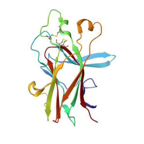
Ligand recognition by A-class Eph receptors: crystal structures of the EphA2 ligand-binding domain and the EphA2/ephrin-A1 complex.

Himanen, J.P.Goldgur, Y.Miao, H.Myshkin, E.Guo, H.Buck, M.Nguyen, M.Rajashankar, K.R.Wang, B.Nikolov, D.B.

(2009) EMBO Rep 10: 722-728

  • DOI: https://doi.org/10.1038/embor.2009.91
  • Primary Citation of Related Structures:  
    3HEI, 3HPN

  • PubMed Abstract: 

    Ephrin (Eph) receptor tyrosine kinases fall into two subclasses (A and B) according to preferences for their ephrin ligands. All published structural studies of Eph receptor/ephrin complexes involve B-class receptors. Here, we present the crystal structures of an A-class complex between EphA2 and ephrin-A1 and of unbound EphA2. Although these structures are similar overall to their B-class counterparts, they reveal important differences that define subclass specificity. The structures suggest that the A-class Eph receptor/ephrin interactions involve smaller rearrangements in the interacting partners, better described by a 'lock-and-key'-type binding mechanism, in contrast to the 'induced fit' mechanism defining the B-class molecules. This model is supported by structure-based mutagenesis and by differential requirements for ligand oligomerization by the two subclasses in cell-based Eph receptor activation assays. Finally, the structure of the unligated receptor reveals a homodimer assembly that might represent EphA2-specific homotypic cell adhesion interactions.


  • Organizational Affiliation

    Structural Biology Program, Memorial Sloan-Kettering Cancer Center, New York, New York 10021, USA.


Macromolecules
Find similar proteins by:  (by identity cutoff)  |  3D Structure
Entity ID: 1
MoleculeChains Sequence LengthOrganismDetailsImage
Ephrin type-A receptor 2
A, B, C, D, E
A, B, C, D, E, F
174Homo sapiensMutation(s): 0 
Gene Names: EPHA2ECK
EC: 2.7.10.1
UniProt & NIH Common Fund Data Resources
Find proteins for P29317 (Homo sapiens)
Explore P29317 
Go to UniProtKB:  P29317
PHAROS:  P29317
GTEx:  ENSG00000142627 
Entity Groups  
Sequence Clusters30% Identity50% Identity70% Identity90% Identity95% Identity100% Identity
UniProt GroupP29317
Sequence Annotations
Expand
  • Reference Sequence
Experimental Data & Validation

Experimental Data

  • Method: X-RAY DIFFRACTION
  • Resolution: 2.52 Å
  • R-Value Free: 0.307 
  • R-Value Work: 0.232 
  • R-Value Observed: 0.236 
  • Space Group: P 1
Unit Cell:
Length ( Å )Angle ( ˚ )
a = 41.271α = 117.14
b = 91.187β = 97.27
c = 91.132γ = 100.79
Software Package:
Software NamePurpose
REFMACrefinement
PDB_EXTRACTdata extraction

Structure Validation

View Full Validation Report


Entry History 

Deposition Data

Revision History  (Full details and data files)

  • Version 1.0: 2009-06-30
    Type: Initial release
  • Version 1.1: 2011-07-13
    Changes: Advisory, Derived calculations, Version format compliance
  • Version 1.2: 2021-03-31
    Changes: Source and taxonomy
  • Version 1.3: 2023-09-06
    Changes: Data collection, Database references, Refinement description