3HIP

HIGH-POTENTIAL IRON-SULFUR PROTEIN FROM CHROMATIUM PURPURATUM


Experimental Data Snapshot

  • Method: X-RAY DIFFRACTION
  • Resolution: 2.80 Å
  • R-Value Free: 0.288 
  • R-Value Work: 0.224 
  • R-Value Observed: 0.224 

wwPDB Validation   3D Report Full Report


This is version 1.3 of the entry. See complete history


Literature

Crystal structure and possible dimerization of the high-potential iron-sulfur protein from Chromatium purpuratum.

Kerfeld, C.A.Salmeen, A.E.Yeates, T.O.

(1998) Biochemistry 37: 13911-13917

  • DOI: https://doi.org/10.1021/bi9810252
  • Primary Citation of Related Structures:  
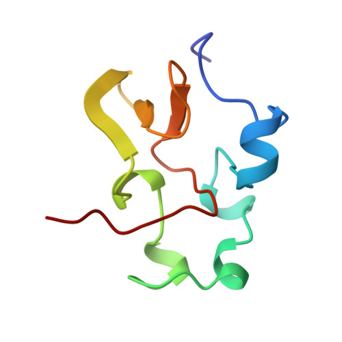
    3HIP

  • PubMed Abstract: 

    The crystal structure of the high-potential iron-sulfur protein (HiPIP) isolated from Chromatium purpuratum is reported at 2.7 A resolution. The three HiPIP molecules in the asymmetric unit of the crystals form one and one-half dimers. Two molecules are related by a noncrystallographic symmetry rotation of approximately 175 degrees with negligible translation along the dyad axis. The third molecule in the asymmetric unit also forms a dimer with a second HiPIP molecule across the crystallographic 2-fold symmetry axis. The Fe4S4 clusters in both the crystallographic and noncrystallographic dimers are separated by approximately 13.0 A. Solution studies give mixed results regarding the oligomeric state of the C. purpuratum HiPIP. A comparison with crystal structures of HiPIPs from other species shows that HiPIP tends to associate rather nonspecifically about a conserved, relatively hydrophobic surface patch to form dimers.


  • Organizational Affiliation

    Molecular Biology Institute, University of California at Los Angeles 90095-1570, USA.


Macromolecules
Find similar proteins by:  (by identity cutoff)  |  3D Structure
Entity ID: 1
MoleculeChains Sequence LengthOrganismDetailsImage
HIGH-POTENTIAL IRON-SULFUR PROTEIN
A, B, C
82Marichromatium purpuratumMutation(s): 0 
UniProt
Find proteins for P59860 (Marichromatium purpuratum)
Explore P59860 
Go to UniProtKB:  P59860
Entity Groups  
Sequence Clusters30% Identity50% Identity70% Identity90% Identity95% Identity100% Identity
UniProt GroupP59860
Sequence Annotations
Expand
  • Reference Sequence
Small Molecules
Experimental Data & Validation

Experimental Data

  • Method: X-RAY DIFFRACTION
  • Resolution: 2.80 Å
  • R-Value Free: 0.288 
  • R-Value Work: 0.224 
  • R-Value Observed: 0.224 
  • Space Group: P 21 21 2
Unit Cell:
Length ( Å )Angle ( ˚ )
a = 55.5α = 90
b = 108.8β = 90
c = 37.2γ = 90
Software Package:
Software NamePurpose
X-PLORmodel building
X-PLORrefinement
DENZOdata reduction
SCALEPACKdata scaling
X-PLORphasing

Structure Validation

View Full Validation Report


Entry History 

Deposition Data

Revision History  (Full details and data files)

  • Version 1.0: 1998-11-11
    Type: Initial release
  • Version 1.1: 2008-03-25
    Changes: Version format compliance
  • Version 1.2: 2011-07-13
    Changes: Version format compliance
  • Version 1.3: 2023-08-09
    Changes: Database references, Derived calculations, Other, Refinement description