3H87

Rv0301 Rv0300 Toxin Antitoxin Complex from Mycobacterium tuberculosis


Experimental Data Snapshot

  • Method: X-RAY DIFFRACTION
  • Resolution: 1.49 Å
  • R-Value Free: 0.174 
  • R-Value Work: 0.156 
  • R-Value Observed: 0.157 

wwPDB Validation   3D Report Full Report


This is version 1.4 of the entry. See complete history


Literature

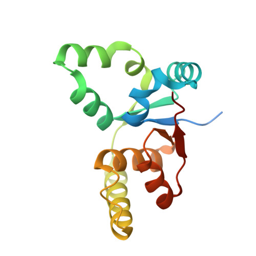
The crystal structure of the Rv0301-Rv0300 VapBC-3 toxin-antitoxin complex from M. tuberculosis reveals a Mg(2+) ion in the active site and a putative RNA-binding site.

Min, A.B.Miallau, L.Sawaya, M.R.Habel, J.Cascio, D.Eisenberg, D.

(2012) Protein Sci 21: 1754-1767

  • DOI: https://doi.org/10.1002/pro.2161
  • Primary Citation of Related Structures:  
    3H87

  • PubMed Abstract: 

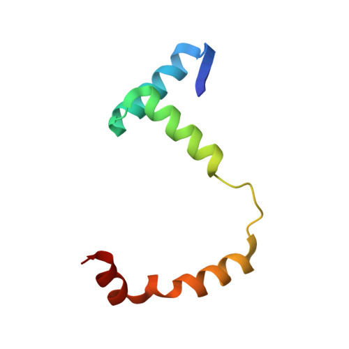
    VapBC pairs account for 45 out of 88 identified toxin-antitoxin (TA) pairs in the Mycobacterium tuberculosis (Mtb) H37Rv genome. A working model suggests that under times of stress, antitoxin molecules are degraded, releasing the toxins to slow the metabolism of the cell, which in the case of VapC toxins is via their RNase activity. Otherwise the TA pairs remain bound to their promoters, autoinhibiting transcription. The crystal structure of Rv0301-Rv0300, an Mtb VapBC TA complex determined at 1.49 Å resolution, suggests a mechanism for these three functions: RNase activity, its inhibition by antitoxin, and its ability to bind promoter DNA. The Rv0301 toxin consists of a core of five parallel beta strands flanked by alpha helices. Three proximal aspartates coordinate a Mg²⁺ ion forming the putative RNase active site. The Rv0300 antitoxin monomer is extended in structure, consisting of an N-terminal beta strand followed by four helices. The last two helices wrap around the toxin and terminate near the putative RNase active site, but with different conformations. In one conformation, the C-terminal arginine interferes with Mg²⁺ ion coordination, suggesting a mechanism by which the antitoxin can inhibit toxin activity. At the N-terminus of the antitoxin, two pairs of Ribbon-Helix-Helix (RHH) motifs are related by crystallographic twofold symmetry. The resulting hetero-octameric complex is similar to the FitAB system, but the two RHH motifs are about 30 Å closer together in the Rv0301-Rv0300 complex, suggesting either a different span of the DNA recognition sequence or a conformational change.


  • Organizational Affiliation

    Department of Chemistry and Biochemistry, Howard Hughes Medical Institute, UCLA-DOE Institute of Genomics and Proteomics, UCLA, Los Angeles, California 90095-1570, USA.


Macromolecules
Find similar proteins by:  (by identity cutoff)  |  3D Structure
Entity ID: 1
MoleculeChains Sequence LengthOrganismDetailsImage
Putative uncharacterized protein
A, B
156Mycobacterium tuberculosis H37RvMutation(s): 0 
Gene Names: MT0314Rv0301
UniProt
Find proteins for P9WFB9 (Mycobacterium tuberculosis (strain ATCC 25618 / H37Rv))
Explore P9WFB9 
Go to UniProtKB:  P9WFB9
Entity Groups  
Sequence Clusters30% Identity50% Identity70% Identity90% Identity95% Identity100% Identity
UniProt GroupP9WFB9
Sequence Annotations
Expand
  • Reference Sequence
Find similar proteins by:  (by identity cutoff)  |  3D Structure
Entity ID: 2
MoleculeChains Sequence LengthOrganismDetailsImage
Putative uncharacterized protein
C, D
73Mycobacterium tuberculosis H37RvMutation(s): 0 
Gene Names: Rv0300
UniProt
Find proteins for O07227 (Mycobacterium tuberculosis (strain ATCC 25618 / H37Rv))
Explore O07227 
Go to UniProtKB:  O07227
Entity Groups  
Sequence Clusters30% Identity50% Identity70% Identity90% Identity95% Identity100% Identity
UniProt GroupO07227
Sequence Annotations
Expand
  • Reference Sequence
Small Molecules
Ligands 4 Unique
IDChains Name / Formula / InChI Key2D Diagram3D Interactions
GOL
Query on GOL

Download Ideal Coordinates CCD File 
E [auth A],
F [auth A],
H [auth B],
I [auth B],
L [auth C]
GLYCEROL
C3 H8 O3
PEDCQBHIVMGVHV-UHFFFAOYSA-N
BME
Query on BME

Download Ideal Coordinates CCD File 
J [auth B]BETA-MERCAPTOETHANOL
C2 H6 O S
DGVVWUTYPXICAM-UHFFFAOYSA-N
IMD
Query on IMD

Download Ideal Coordinates CCD File 
K [auth B]IMIDAZOLE
C3 H5 N2
RAXXELZNTBOGNW-UHFFFAOYSA-O
MG
Query on MG

Download Ideal Coordinates CCD File 
G [auth A]MAGNESIUM ION
Mg
JLVVSXFLKOJNIY-UHFFFAOYSA-N
Experimental Data & Validation

Experimental Data

  • Method: X-RAY DIFFRACTION
  • Resolution: 1.49 Å
  • R-Value Free: 0.174 
  • R-Value Work: 0.156 
  • R-Value Observed: 0.157 
  • Space Group: P 41 21 2
Unit Cell:
Length ( Å )Angle ( ˚ )
a = 85.557α = 90
b = 85.557β = 90
c = 155.611γ = 90
Software Package:
Software NamePurpose
DENZOdata reduction
SCALEPACKdata scaling
SHELXphasing
DMphasing
REFMACrefinement
PDB_EXTRACTdata extraction
ADSCdata collection
SHELXDphasing

Structure Validation

View Full Validation Report


Entry History 

Deposition Data

Revision History  (Full details and data files)

  • Version 1.0: 2009-05-05
    Type: Initial release
  • Version 1.1: 2011-07-13
    Changes: Advisory, Version format compliance
  • Version 1.2: 2012-11-07
    Changes: Database references
  • Version 1.3: 2017-11-01
    Changes: Refinement description
  • Version 1.4: 2024-02-21
    Changes: Data collection, Database references, Derived calculations