3GOV

Crystal structure of the catalytic region of human MASP-1


Experimental Data Snapshot

  • Method: X-RAY DIFFRACTION
  • Resolution: 2.55 Å
  • R-Value Free: 0.278 
  • R-Value Work: 0.229 
  • R-Value Observed: 0.231 

wwPDB Validation   3D Report Full Report


This is version 1.3 of the entry. See complete history


Literature

MASP-1, a promiscuous complement protease: structure of its catalytic region reveals the basis of its broad specificity.

Dobo, J.Harmat, V.Beinrohr, L.Sebestyen, E.Zavodszky, P.Gal, P.

(2009) J Immunol 183: 1207-1214

  • DOI: https://doi.org/10.4049/jimmunol.0901141
  • Primary Citation of Related Structures:  
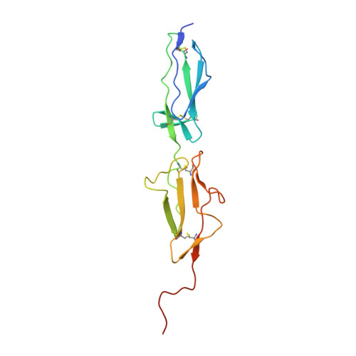
    3GOV

  • PubMed Abstract: 

    Mannose-binding lectin (MBL)-associated serine protease (MASP)-1 is an abundant component of the lectin pathway of complement. The related enzyme, MASP-2 is capable of activating the complement cascade alone. Though the concentration of MASP-1 far exceeds that of MASP-2, only a supporting role of MASP-1 has been identified regarding lectin pathway activation. Several non-complement substrates, like fibrinogen and factor XIII, have also been reported. MASP-1 belongs to the C1r/C1s/MASP family of modular serine proteases; however, its serine protease domain is evolutionary different. We have determined the crystal structure of the catalytic region of active MASP-1 and refined it to 2.55 A resolution. Unusual features of the structure are an internal salt bridge (similar to one in factor D) between the S1 Asp189 and Arg224, and a very long 60-loop. The functional and evolutionary differences between MASP-1 and the other members of the C1r/C1s/MASP family are reflected in the crystal structure. Structural comparison of the protease domains revealed that the substrate binding groove of MASP-1 is wide and resembles that of trypsin rather than early complement proteases explaining its relaxed specificity. Also, MASP-1's multifunctional behavior as both a complement and a coagulation enzyme is in accordance with our observation that antithrombin in the presence of heparin is a more potent inhibitor of MASP-1 than C1 inhibitor. Overall, MASP-1 behaves as a promiscuous protease. The structure shows that its substrate binding groove is accessible; however, its reactivity could be modulated by an unusually large 60-loop and an internal salt bridge involving the S1 Asp.


  • Organizational Affiliation

    Institute of Enzymology, Biological Research Center, Hungarian Academy of Sciences, Budapest, Hungary. dobo@enzim.hu


Macromolecules
Find similar proteins by:  (by identity cutoff)  |  3D Structure
Entity ID: 1
MoleculeChains Sequence LengthOrganismDetailsImage
MASP-1155Homo sapiensMutation(s): 0 
Gene Names: CRARFCRARF1MASP1PRSS5
EC: 3.4.21
UniProt & NIH Common Fund Data Resources
Find proteins for P48740 (Homo sapiens)
Explore P48740 
Go to UniProtKB:  P48740
PHAROS:  P48740
GTEx:  ENSG00000127241 
Entity Groups  
Sequence Clusters30% Identity50% Identity70% Identity90% Identity95% Identity100% Identity
UniProt GroupP48740
Sequence Annotations
Expand
  • Reference Sequence
Find similar proteins by:  (by identity cutoff)  |  3D Structure
Entity ID: 2
MoleculeChains Sequence LengthOrganismDetailsImage
MASP-1251Homo sapiensMutation(s): 0 
Gene Names: CRARFCRARF1MASP1PRSS5
EC: 3.4.21
UniProt & NIH Common Fund Data Resources
Find proteins for P48740 (Homo sapiens)
Explore P48740 
Go to UniProtKB:  P48740
PHAROS:  P48740
GTEx:  ENSG00000127241 
Entity Groups  
Sequence Clusters30% Identity50% Identity70% Identity90% Identity95% Identity100% Identity
UniProt GroupP48740
Sequence Annotations
Expand
  • Reference Sequence
Experimental Data & Validation

Experimental Data

  • Method: X-RAY DIFFRACTION
  • Resolution: 2.55 Å
  • R-Value Free: 0.278 
  • R-Value Work: 0.229 
  • R-Value Observed: 0.231 
  • Space Group: P 21 21 21
Unit Cell:
Length ( Å )Angle ( ˚ )
a = 68.418α = 90
b = 70.412β = 90
c = 121.413γ = 90
Software Package:
Software NamePurpose
XSCALEdata scaling
MOLREPphasing
REFMACrefinement
PDB_EXTRACTdata extraction
MAR345dtbdata collection
XDSdata reduction

Structure Validation

View Full Validation Report


Entry History 

Deposition Data

Revision History  (Full details and data files)

  • Version 1.0: 2009-06-09
    Type: Initial release
  • Version 1.1: 2011-07-13
    Changes: Advisory, Version format compliance
  • Version 1.2: 2013-09-25
    Changes: Derived calculations
  • Version 1.3: 2023-09-06
    Changes: Data collection, Database references, Derived calculations, Refinement description