3GIT

Crystal structure of a truncated acetyl-CoA synthase


Experimental Data Snapshot

  • Method: X-RAY DIFFRACTION
  • Resolution: 3.00 Å
  • R-Value Free: 0.208 
  • R-Value Work: 0.171 
  • R-Value Observed: 0.173 

wwPDB Validation   3D Report Full Report


This is version 1.2 of the entry. See complete history


Literature

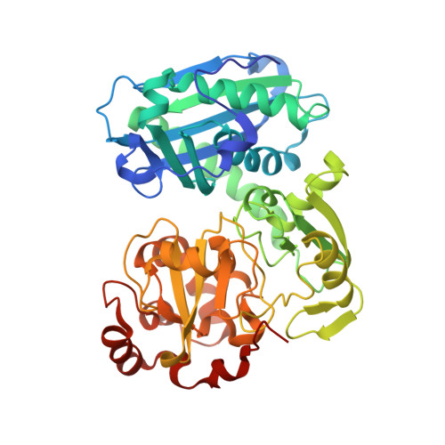
Novel domain arrangement in the crystal structure of a truncated acetyl-CoA synthase from Moorella thermoacetica

Volbeda, A.Darnault, C.Tan, X.Lindahl, P.A.Fontecilla-Camps, J.C.

(2009) Biochemistry 48: 7916-7926

  • DOI: https://doi.org/10.1021/bi9003952
  • Primary Citation of Related Structures:  
    3GIT

  • PubMed Abstract: 

    Ni-dependent acetyl-CoA synthase (ACS) and CO dehydrogenase (CODH) constitute the central enzyme complex of the Wood-Ljungdahl pathway of acetyl-CoA formation. The crystal structure of a recombinant bacterial ACS lacking the N-terminal domain that interacts with CODH shows a large reorganization of the remaining two globular domains, producing a narrow cleft of suitable size, shape, and nature to bind CoA. Sequence comparisons with homologous archaeal enzymes that naturally lack the N-terminal domain show that many amino acids lining this cleft are conserved. Besides the typical [4Fe-4S] center, the A-cluster contains only one proximal metal ion that, according to anomalous scattering data, is most likely Cu or Zn. Incorporation of a functional Ni(2)Fe(4)S(4) A-cluster would require only minor structural rearrangements. Using available structures, a plausible model of the interaction between CODH and the smaller ACS in archaeal multienzyme complexes is presented, along with a discussion of evolutionary relationships of the archaeal and bacterial enzymes.


  • Organizational Affiliation

    Laboratoire de Cristallographie et Cristallogenèse des Protéines, Institut de Biologie Structurale Jean-Pierre Ebel, CEA, CNRS, Université Joseph Fourier, 41 Rue Jules Horowitz, F-38027 Grenoble, France. anne.volbeda@ibs.fr


Macromolecules
Find similar proteins by:  (by identity cutoff)  |  3D Structure
Entity ID: 1
MoleculeChains Sequence LengthOrganismDetailsImage
Carbon monoxide dehydrogenase/acetyl-CoA synthase subunit alpha
A, B, C, D, E
A, B, C, D, E, F
427Moorella thermoaceticaMutation(s): 0 
Gene Names: acsB2
EC: 2.3.1.169
UniProt
Find proteins for P27988 (Moorella thermoacetica)
Explore P27988 
Go to UniProtKB:  P27988
Entity Groups  
Sequence Clusters30% Identity50% Identity70% Identity90% Identity95% Identity100% Identity
UniProt GroupP27988
Sequence Annotations
Expand
  • Reference Sequence
Small Molecules
Ligands 5 Unique
IDChains Name / Formula / InChI Key2D Diagram3D Interactions
SF4
Query on SF4

Download Ideal Coordinates CCD File 
G [auth A]
GA [auth D]
NA [auth E]
P [auth B]
VA [auth F]
G [auth A],
GA [auth D],
NA [auth E],
P [auth B],
VA [auth F],
X [auth C]
IRON/SULFUR CLUSTER
Fe4 S4
LJBDFODJNLIPKO-UHFFFAOYSA-N
SO4
Query on SO4

Download Ideal Coordinates CCD File 
AA [auth C]
AB [auth F]
BB [auth F]
CA [auth C]
DA [auth C]
AA [auth C],
AB [auth F],
BB [auth F],
CA [auth C],
DA [auth C],
EA [auth C],
FA [auth D],
J [auth A],
JA [auth D],
K [auth A],
LA [auth D],
M [auth A],
MA [auth D],
N [auth A],
O [auth A],
QA [auth E],
S [auth B],
SA [auth E],
TA [auth E],
U [auth B],
UA [auth F],
V [auth B],
W [auth B],
YA [auth F]
SULFATE ION
O4 S
QAOWNCQODCNURD-UHFFFAOYSA-L
GOL
Query on GOL

Download Ideal Coordinates CCD File 
BA [auth C]
KA [auth D]
L [auth A]
RA [auth E]
T [auth B]
BA [auth C],
KA [auth D],
L [auth A],
RA [auth E],
T [auth B],
ZA [auth F]
GLYCEROL
C3 H8 O3
PEDCQBHIVMGVHV-UHFFFAOYSA-N
ZN
Query on ZN

Download Ideal Coordinates CCD File 
H [auth A]
HA [auth D]
OA [auth E]
Q [auth B]
WA [auth F]
H [auth A],
HA [auth D],
OA [auth E],
Q [auth B],
WA [auth F],
Y [auth C]
ZINC ION
Zn
PTFCDOFLOPIGGS-UHFFFAOYSA-N
H2S
Query on H2S

Download Ideal Coordinates CCD File 
I [auth A]
IA [auth D]
PA [auth E]
R [auth B]
XA [auth F]
I [auth A],
IA [auth D],
PA [auth E],
R [auth B],
XA [auth F],
Z [auth C]
HYDROSULFURIC ACID
H2 S
RWSOTUBLDIXVET-UHFFFAOYSA-N
Experimental Data & Validation

Experimental Data

  • Method: X-RAY DIFFRACTION
  • Resolution: 3.00 Å
  • R-Value Free: 0.208 
  • R-Value Work: 0.171 
  • R-Value Observed: 0.173 
  • Space Group: P 31 2 1
Unit Cell:
Length ( Å )Angle ( ˚ )
a = 166.4α = 90
b = 166.4β = 90
c = 245.2γ = 120
Software Package:
Software NamePurpose
XSCALEdata scaling
PHASERphasing
REFMACrefinement
PDB_EXTRACTdata extraction
Xnemodata collection
XDSdata reduction

Structure Validation

View Full Validation Report


Entry History 

Deposition Data

Revision History  (Full details and data files)

  • Version 1.0: 2009-10-06
    Type: Initial release
  • Version 1.1: 2011-07-13
    Changes: Advisory, Version format compliance
  • Version 1.2: 2023-11-01
    Changes: Data collection, Database references, Derived calculations, Refinement description