3GFS

Structure of YhdA, K109D/D137K variant


Experimental Data Snapshot

  • Method: X-RAY DIFFRACTION
  • Resolution: 2.10 Å
  • R-Value Free: 0.221 
  • R-Value Work: 0.185 
  • R-Value Observed: 0.187 

wwPDB Validation   3D Report Full Report


Ligand Structure Quality Assessment 


This is version 1.3 of the entry. See complete history


Literature

A single intersubunit salt bridge affects oligomerization and catalytic activity in a bacterial quinone reductase

Binter, A.Staunig, N.Jelesarov, I.Lohner, K.Palfey, B.A.Deller, S.Gruber, K.Macheroux, P.

(2009) FEBS J 276: 5263-5274

  • DOI: https://doi.org/10.1111/j.1742-4658.2009.07222.x
  • Primary Citation of Related Structures:  
    3GFQ, 3GFR, 3GFS

  • PubMed Abstract: 

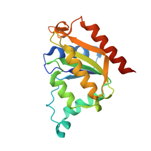
    YhdA, a thermostable NADPH:FMN oxidoreductase from Bacillus subtilis, reduces quinones via a ping-pong bi-bi mechanism with a pronounced preference for NADPH. The enzyme occurs as a stable tetramer in solution. The two extended dimer surfaces are packed against each other by a 90 rotation of one dimer with respect to the other. This assembly is stabilized by the formation of four salt bridges between K109 and D137 of the neighbouring protomers. To investigate the importance of the ion pair contacts, the K109L and D137L single replacement variants, as well as the K109L/D137L and K109D/D137K double replacement variants, were generated, expressed, purified, crystallized and biochemically characterized. The K109L and D137L variants form dimers instead of tetramers, whereas the K109L/D137L and K109D/D137K variants appear to exist in a dimer-tetramer equilibrium in solution. The crystal structures of the K109L and D137L variants confirm the dimeric state, with the K109L/D137L and K109D/D137K variants adopting a tetrameric assembly. Interestingly, all protein variants show a drastically reduced quinone reductase activity in steady-state kinetics. Detailed analysis of the two half reactions revealed that the oxidative half reaction is not affected, whereas reduction of the bound FMN cofactor by NADPH is virtually abolished. Inspection of the crystal structures indicates that the side chain of K109 plays a dual role by forming a salt bridge to D137, as well as stabilizing a glycine-rich loop in the vicinity of the FMN cofactor. In all protein variants, this glycine-rich loop exhibits a much higher mobility, compared to the wild-type. This appears to be incompatible with NADPH binding and thus leads to abrogation of flavin reduction.


  • Organizational Affiliation

    Institute of Biochemistry, Graz University of Technology, Austria.


Macromolecules
Find similar proteins by:  (by identity cutoff)  |  3D Structure
Entity ID: 1
MoleculeChains Sequence LengthOrganismDetailsImage
FMN-dependent NADPH-azoreductase
A, B, C, D, E
A, B, C, D, E, F, G, H, I, J, K, L
174Bacillus subtilisMutation(s): 2 
Gene Names: azrBSU09340yhdA
EC: 1.7
UniProt
Find proteins for O07529 (Bacillus subtilis (strain 168))
Explore O07529 
Go to UniProtKB:  O07529
Entity Groups  
Sequence Clusters30% Identity50% Identity70% Identity90% Identity95% Identity100% Identity
UniProt GroupO07529
Sequence Annotations
Expand
  • Reference Sequence
Small Molecules
Ligands 1 Unique
IDChains Name / Formula / InChI Key2D Diagram3D Interactions
FMN
Query on FMN

Download Ideal Coordinates CCD File 
M [auth A]
N [auth B]
O [auth C]
P [auth D]
Q [auth E]
M [auth A],
N [auth B],
O [auth C],
P [auth D],
Q [auth E],
R [auth F],
S [auth G],
T [auth H],
U [auth I],
V [auth J],
W [auth K],
X [auth L]
FLAVIN MONONUCLEOTIDE
C17 H21 N4 O9 P
FVTCRASFADXXNN-SCRDCRAPSA-N
Experimental Data & Validation

Experimental Data

  • Method: X-RAY DIFFRACTION
  • Resolution: 2.10 Å
  • R-Value Free: 0.221 
  • R-Value Work: 0.185 
  • R-Value Observed: 0.187 
  • Space Group: P 1 21 1
Unit Cell:
Length ( Å )Angle ( ˚ )
a = 68.625α = 90
b = 170.132β = 92.25
c = 93.289γ = 90
Software Package:
Software NamePurpose
PHENIXrefinement
PDB_EXTRACTdata extraction
MAR345dtbdata collection
DENZOdata reduction
SCALEPACKdata scaling
PHASERphasing

Structure Validation

View Full Validation Report


Ligand Structure Quality Assessment 


Entry History 

Deposition Data

Revision History  (Full details and data files)

  • Version 1.0: 2009-09-22
    Type: Initial release
  • Version 1.1: 2011-07-13
    Changes: Version format compliance
  • Version 1.2: 2021-10-20
    Changes: Data collection, Database references, Derived calculations
  • Version 1.3: 2023-09-06
    Changes: Data collection, Refinement description