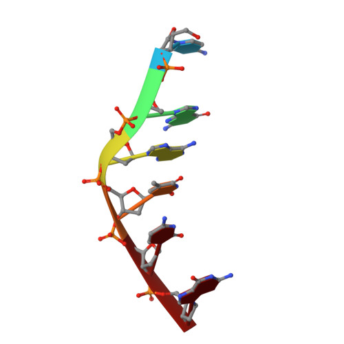
Crystal Structure of Proflavine in Complex with a DNA hexamer duplex
Maehigashi, T., Persil, O., Hud, N.V., Williams, L.D.To be published.
Experimental Data Snapshot
| Ligands 2 Unique | |||||
|---|---|---|---|---|---|
| ID | Chains | Name / Formula / InChI Key | 2D Diagram | 3D Interactions | |
| PRL Query on PRL | C [auth A], F [auth B] | PROFLAVIN C13 H11 N3 WDVSHHCDHLJJJR-UHFFFAOYSA-N |  | ||
| NCO Query on NCO | D [auth A], E [auth A], G [auth B] | COBALT HEXAMMINE(III) Co H18 N6 DYLMFCCYOUSRTK-UHFFFAOYSA-N |  | ||
| Length ( Å ) | Angle ( ˚ ) |
|---|---|
| a = 24.563 | α = 90 |
| b = 24.563 | β = 90 |
| c = 78.276 | γ = 120 |
| Software Name | Purpose |
|---|---|
| REFMAC | refinement |
| CNS | refinement |
| CBASS | data collection |
| HKL-2000 | data reduction |
| HKL-2000 | data scaling |
| CNS | phasing |
RCSB PDB (citation) is hosted by
RCSB PDB is a member of the