3FOI

Fitting of gp18M crystal structure into 3D cryo-EM reconstruction of bacteriophage T4 contracted tail


Experimental Data Snapshot

  • Method: ELECTRON MICROSCOPY
  • Resolution: 16.0 Å
  • Aggregation State: PARTICLE 
  • Reconstruction Method: SINGLE PARTICLE 

wwPDB Validation   3D Report Full Report


This is version 1.6 of the entry. See complete history


Literature

The tail sheath structure of bacteriophage T4: a molecular machine for infecting bacteria.

Aksyuk, A.A.Leiman, P.G.Kurochkina, L.P.Shneider, M.M.Kostyuchenko, V.A.Mesyanzhinov, V.V.Rossmann, M.G.

(2009) EMBO J 28: 821-829

  • DOI: https://doi.org/10.1038/emboj.2009.36
  • Primary Citation of Related Structures:  
    3FO8, 3FOA, 3FOH, 3FOI

  • PubMed Abstract: 

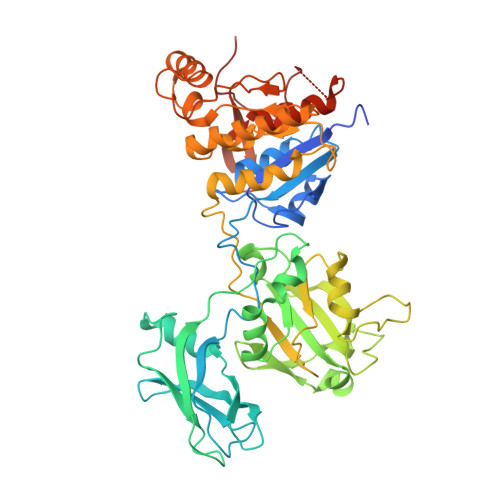
    The contractile tail of bacteriophage T4 is a molecular machine that facilitates very high viral infection efficiency. Its major component is a tail sheath, which contracts during infection to less than half of its initial length. The sheath consists of 138 copies of the tail sheath protein, gene product (gp) 18, which surrounds the central non-contractile tail tube. The contraction of the sheath drives the tail tube through the outer membrane, creating a channel for the viral genome delivery. A crystal structure of about three quarters of gp18 has been determined and was fitted into cryo-electron microscopy reconstructions of the tail sheath before and after contraction. It was shown that during contraction, gp18 subunits slide over each other with no apparent change in their structure.


  • Organizational Affiliation

    Department of Biological Sciences, Purdue University, West Lafayette, IN 47907-2054, USA.


Macromolecules
Find similar proteins by:  (by identity cutoff)  |  3D Structure
Entity ID: 1
MoleculeChains Sequence LengthOrganismDetailsImage
Tail sheath protein Gp18
A, B, C, D, E
A, B, C, D, E, F
510Tequatrovirus T4Mutation(s): 1 
Gene Names: 18GB AAA32541
UniProt
Find proteins for P13332 (Enterobacteria phage T4)
Explore P13332 
Go to UniProtKB:  P13332
Entity Groups  
Sequence Clusters30% Identity50% Identity70% Identity90% Identity95% Identity100% Identity
UniProt GroupP13332
Sequence Annotations
Expand
  • Reference Sequence
Experimental Data & Validation

Experimental Data

  • Method: ELECTRON MICROSCOPY
  • Resolution: 16.0 Å
  • Aggregation State: PARTICLE 
  • Reconstruction Method: SINGLE PARTICLE 
EM Software:
TaskSoftware PackageVersion
RECONSTRUCTIONSPIDER

Structure Validation

View Full Validation Report


Entry History 

Deposition Data

Revision History  (Full details and data files)

  • Version 1.0: 2009-03-10
    Type: Initial release
  • Version 1.1: 2011-07-13
    Changes: Version format compliance
  • Version 1.2: 2012-05-30
    Changes: Refinement description
  • Version 1.3: 2018-07-18
    Changes: Data collection
  • Version 1.4: 2019-11-06
    Changes: Data collection, Other
  • Version 1.5: 2021-10-20
    Changes: Database references
  • Version 1.6: 2024-02-21
    Changes: Data collection, Refinement description