3FDO

Structure of human MDMX in complex with high affinity peptide


Experimental Data Snapshot

  • Method: X-RAY DIFFRACTION
  • Resolution: 1.40 Å
  • R-Value Free: 0.247 
  • R-Value Work: 0.179 
  • R-Value Observed: 0.183 

wwPDB Validation   3D Report Full Report


This is version 1.3 of the entry. See complete history


Literature

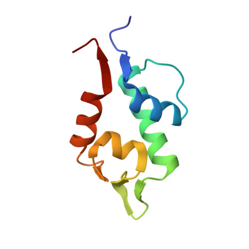
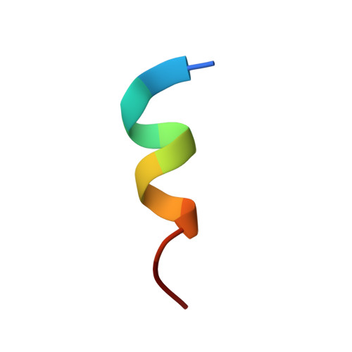
High affinity interaction of the p53 peptide-analogue with human Mdm2 and Mdmx.

Czarna, A.Popowicz, G.M.Pecak, A.Wolf, S.Dubin, G.Holak, T.A.

(2009) Cell Cycle 8: 1176-1184

  • DOI: https://doi.org/10.4161/cc.8.8.8185
  • Primary Citation of Related Structures:  
    3FDO, 3G03

  • PubMed Abstract: 

    The Mdm2 and Mdmx proteins are the principal negative regulators of the p53 tumor suppressor. Reactivation of p53 activity by disrupting the Mdm2/Mdmx-p53 interactions offers new possibilities for anticancer therapeutics. Here, we present crystal structures of two complexes, a p53-like mutant peptide with the N-terminal domains of Mdm2 and Mdmx, respectively. The structures reveal that the p53 mutant peptide (amino acid sequence: LTFEHYWAQLTS) assumes virtually identical conformations in both complexes despite the different shapes of the p53-binding pockets in these two proteins, has a more extended helical nature compared to the Mdm2-bound wild-type p53 peptide, and does not disturb the native folds of Mdm2 or Mdmx. The extension of the helical structure in the mutant p53 peptide greatly improves its binding to Mdm2 and Mdmx. The fluorescence polarization assay that we have developed using this peptide indicates the affinities towards Mdm2 of 3.6 nM and for Mdmx of 6.1 nM, compared to the low micromolar binding of a similar length wild-type p53 peptide to Mdm2/Mdmx. Our assay does not require expensive non-native amino acids, and allows measurements of the interaction with both Mdm2 and Mdmx in identical conditions-without modification of experimental conditions or setups between the two proteins. The structural information presented here, coupled with the robust fluorescence polarization assay, should enable development of a simple pharmacophore model of cross-selective Mdm2-Mdmx/p53 inhibitors.


  • Organizational Affiliation

    Max Planck Institute for Biochemistry, Martinsried, Germany.


Macromolecules
Find similar proteins by:  (by identity cutoff)  |  3D Structure
Entity ID: 1
MoleculeChains Sequence LengthOrganismDetailsImage
Protein Mdm490Homo sapiensMutation(s): 0 
Gene Names: MDM4MDMX
UniProt & NIH Common Fund Data Resources
Find proteins for O15151 (Homo sapiens)
Explore O15151 
Go to UniProtKB:  O15151
PHAROS:  O15151
GTEx:  ENSG00000198625 
Entity Groups  
Sequence Clusters30% Identity50% Identity70% Identity90% Identity95% Identity100% Identity
UniProt GroupO15151
Sequence Annotations
Expand
  • Reference Sequence

Find similar proteins by:  Sequence   |   3D Structure  

Entity ID: 2
MoleculeChains Sequence LengthOrganismDetailsImage
Synthetic high affinity peptide12N/AMutation(s): 0 
Entity Groups  
Sequence Clusters30% Identity50% Identity70% Identity90% Identity95% Identity100% Identity
Sequence Annotations
Expand
  • Reference Sequence
Experimental Data & Validation

Experimental Data

  • Method: X-RAY DIFFRACTION
  • Resolution: 1.40 Å
  • R-Value Free: 0.247 
  • R-Value Work: 0.179 
  • R-Value Observed: 0.183 
  • Space Group: C 1 2 1
Unit Cell:
Length ( Å )Angle ( ˚ )
a = 69.23α = 90
b = 30.95β = 124.41
c = 50.42γ = 90
Software Package:
Software NamePurpose
MAR345dtbdata collection
MOLREPphasing
REFMACrefinement
XDSdata reduction
XSCALEdata scaling

Structure Validation

View Full Validation Report


Entry History 

Deposition Data

Revision History  (Full details and data files)

  • Version 1.0: 2008-12-16
    Type: Initial release
  • Version 1.1: 2011-07-13
    Changes: Derived calculations, Version format compliance
  • Version 1.2: 2019-11-06
    Changes: Data collection, Database references
  • Version 1.3: 2023-11-01
    Changes: Data collection, Database references, Derived calculations, Refinement description