3FB1

Crystal Structure of Purine Nucleoside Phosphorylase in Complex with Ribose-1-Phosphate


Experimental Data Snapshot

  • Method: X-RAY DIFFRACTION
  • Resolution: 2.00 Å
  • R-Value Free: 0.234 
  • R-Value Work: 0.175 
  • R-Value Observed: 0.177 

wwPDB Validation   3D Report Full Report


Ligand Structure Quality Assessment 


This is version 2.1 of the entry. See complete history


Literature

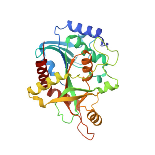
Purine nucleoside phosphorylase from Schistosoma mansoni in complex with ribose-1-phosphate.

D'Muniz Pereira, H.Oliva, G.Garratt, R.C.

(2011) J Synchrotron Radiat 18: 62-65

  • DOI: https://doi.org/10.1107/S0909049510027718
  • Primary Citation of Related Structures:  
    3FB1

  • PubMed Abstract: 

    Schistosomes are blood flukes which cause schistosomiasis, a disease affecting approximately 200 million people worldwide. Along with several other important human parasites including trypanosomes and Plasmodium, schistosomes lack the de novo pathway for purine synthesis and depend exclusively on the salvage pathway for their purine requirements, making the latter an attractive target for drug development. Part of the pathway involves the conversion of inosine (or guanosine) into hypoxanthine (or guanine) together with ribose-1-phosphate (R1P) or vice versa. This inter-conversion is undertaken by the enzyme purine nucleoside phosphorylase (PNP) which has been used as the basis for the development of novel anti-malarials, conceptually validating this approach. It has been suggested that, during the reverse reaction, R1P binding to the enzyme would occur only as a consequence of conformational changes induced by hypoxanthine, thus making a binary PNP-R1P complex unlikely. Contradictory to this statement, a crystal structure of just such a binary complex involving the Schistosoma mansoni enzyme has been successfully obtained. The ligand shows an intricate hydrogen-bonding network in the phosphate and ribose binding sites and adds a further chapter to our knowledge which could be of value in the future development of selective inhibitors.


  • Organizational Affiliation

    Centro de Biotecnologia Molecular Estrutural, Instituto de Física de São Carlos, Universidade de São Paulo, Avenida Trabalhador São-Carlense 400, CEP 13566-590, Brazil. hmuniz.pereira@gmail.com


Macromolecules
Find similar proteins by:  (by identity cutoff)  |  3D Structure
Entity ID: 1
MoleculeChains Sequence LengthOrganismDetailsImage
Purine-nucleoside phosphorylase
A, B, C
287Schistosoma mansoniMutation(s): 0 
Gene Names: SmPNP
EC: 2.4.2.1
UniProt
Find proteins for Q9BMI9 (Schistosoma mansoni)
Explore Q9BMI9 
Go to UniProtKB:  Q9BMI9
Entity Groups  
Sequence Clusters30% Identity50% Identity70% Identity90% Identity95% Identity100% Identity
UniProt GroupQ9BMI9
Sequence Annotations
Expand
  • Reference Sequence
Experimental Data & Validation

Experimental Data

  • Method: X-RAY DIFFRACTION
  • Resolution: 2.00 Å
  • R-Value Free: 0.234 
  • R-Value Work: 0.175 
  • R-Value Observed: 0.177 
  • Space Group: P 21 21 21
Unit Cell:
Length ( Å )Angle ( ˚ )
a = 48.965α = 90
b = 117.669β = 90
c = 129.514γ = 90
Software Package:
Software NamePurpose
SCALAdata scaling
MOLREPphasing
PHENIXrefinement
PDB_EXTRACTdata extraction
MAR345dtbdata collection
MOSFLMdata reduction

Structure Validation

View Full Validation Report


Ligand Structure Quality Assessment 


Entry History 

Deposition Data

Revision History  (Full details and data files)

  • Version 1.0: 2009-11-24
    Type: Initial release
  • Version 1.1: 2011-07-13
    Changes: Version format compliance
  • Version 1.2: 2012-01-25
    Changes: Database references
  • Version 2.0: 2020-07-29
    Type: Remediation
    Reason: Carbohydrate remediation
    Changes: Atomic model, Data collection, Derived calculations, Structure summary
  • Version 2.1: 2023-12-27
    Changes: Data collection, Database references, Structure summary