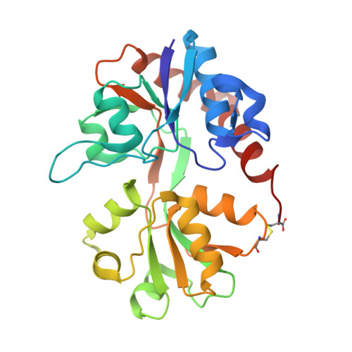
Correlating AMPA receptor activation and cleft closure across subunits: crystal structures of the GluR4 ligand-binding domain in complex with full and partial agonists
Gill, A., Birdsey-Benson, A., Jones, B.L., Henderson, L.P., Madden, D.R.(2008) Biochemistry 47: 13831-13841
- PubMed: 19102704
- DOI: https://doi.org/10.1021/bi8013196
- Primary Citation of Related Structures:
3EN3, 3EPE - PubMed Abstract:
AMPA receptors are glutamate-gated ion channels that are essential mediators of synaptic signals in the central nervous system. They form tetramers that are assembled as combinations of subunits GluR1-4, each of which contains a ligand-binding domain (LBD). Crystal structures of the GluR2 LBD have revealed an agonist-binding cleft, which is located between two lobes and which acts like a Venus flytrap. In general, agonist efficacy is correlated with the extent of cleft closure. However, recent observations show that cleft closure is not the sole determinant of the relative efficacy for glutamate receptors. In addition, these studies have focused on the GluR2 subunit, which is the specific target of a physiologically important RNA-editing modification in vivo. We therefore sought to test the generality of the cleft closure-efficacy correlation for other AMPA-R subunits. Here, we present crystal structures of the GluR4(flip) LBD in complex with both full and partial agonists. As for GluR2, both agonists stabilize a closed-cleft conformation, and the partial agonist induces a smaller cleft closure than the full agonist. However, a detailed analysis of LBD-kainate interactions reveals the importance of subtle backbone conformational changes in the ligand-binding pocket in determining the magnitude of agonist-associated conformational changes. Furthermore, the GluR4 subunit exhibits a different correlation between receptor activation and LBD cleft closure than does GluR2.
Organizational Affiliation:
Departments of Biochemistry and Physiology, Dartmouth Medical School, 7200 Vail Building, Hanover, New Hampshire 03755, USA.