3EE1

Novel fold of VirA, a type III secretion system effector protein from Shigella flexneri


Experimental Data Snapshot

  • Method: X-RAY DIFFRACTION
  • Resolution: 3.01 Å
  • R-Value Free: 0.256 
  • R-Value Work: 0.214 
  • R-Value Observed: 0.216 

wwPDB Validation   3D Report Full Report


This is version 1.2 of the entry. See complete history


Literature

Novel fold of VirA, a type III secretion system effector protein from Shigella flexneri

Davis, J.Wang, J.Tropea, J.E.Zhang, D.Dauter, Z.Waugh, D.S.Wlodawer, A.

(2008) Protein Sci 17: 2167-2173

  • DOI: https://doi.org/10.1110/ps.037978.108
  • Primary Citation of Related Structures:  
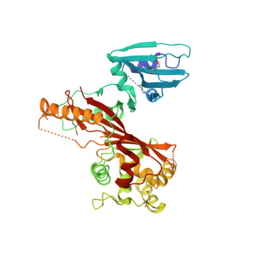
    3EE1

  • PubMed Abstract: 

    VirA, a secreted effector protein from Shigella sp., has been shown to be necessary for its virulence. It was also reported that VirA might be related to papain-like cysteine proteases and cleave alpha-tubulin, thus facilitating intracellular spreading. We have now determined the crystal structure of VirA at 3.0 A resolution. The shape of the molecule resembles the letter "V," with the residues in the N-terminal third of the 45-kDa molecule (some of which are disordered) forming one clearly identifiable domain, and the remainder of the molecule completing the V-like structure. The fold of VirA is unique and does not resemble that of any known protein, including papain, although its N-terminal domain is topologically similar to cysteine protease inhibitors such as stefin B. Analysis of the sequence conservation between VirA and its Escherichia coli homologs EspG and EspG2 did not result in identification of any putative protease-like active site, leaving open a possibility that the biological function of VirA in Shigella virulence may not involve direct proteolytic activity.


  • Organizational Affiliation

    Protein Structure Section, Macromolecular Crystallography Laboratory, NCI, Frederick, Maryland 21702, USA.


Macromolecules
Find similar proteins by:  (by identity cutoff)  |  3D Structure
Entity ID: 1
MoleculeChains Sequence LengthOrganismDetailsImage
Effector protein virA
A, B
400Shigella flexneriMutation(s): 4 
Gene Names: virA
EC: 3.4.22
UniProt
Find proteins for Q7BU69 (Shigella flexneri)
Explore Q7BU69 
Go to UniProtKB:  Q7BU69
Entity Groups  
Sequence Clusters30% Identity50% Identity70% Identity90% Identity95% Identity100% Identity
UniProt GroupQ7BU69
Sequence Annotations
Expand
  • Reference Sequence
Small Molecules
Modified Residues  1 Unique
IDChains TypeFormula2D DiagramParent
MSE
Query on MSE
A, B
L-PEPTIDE LINKINGC5 H11 N O2 SeMET
Experimental Data & Validation

Experimental Data

  • Method: X-RAY DIFFRACTION
  • Resolution: 3.01 Å
  • R-Value Free: 0.256 
  • R-Value Work: 0.214 
  • R-Value Observed: 0.216 
  • Space Group: C 1 2 1
Unit Cell:
Length ( Å )Angle ( ˚ )
a = 150.252α = 90
b = 170.91β = 104.89
c = 46.165γ = 90
Software Package:
Software NamePurpose
DENZOdata reduction
SCALEPACKdata scaling
PHENIXrefinement
PDB_EXTRACTdata extraction

Structure Validation

View Full Validation Report


Entry History 

Deposition Data

  • Released Date: 2008-12-16 
  • Deposition Author(s): Davis, J.S.

Revision History  (Full details and data files)

  • Version 1.0: 2008-12-16
    Type: Initial release
  • Version 1.1: 2011-07-13
    Changes: Version format compliance
  • Version 1.2: 2017-10-25
    Changes: Refinement description