3ECA

CRYSTAL STRUCTURE OF ESCHERICHIA COLI L-ASPARAGINASE, AN ENZYME USED IN CANCER THERAPY (ELSPAR)


Experimental Data Snapshot

  • Method: X-RAY DIFFRACTION
  • Resolution: 2.40 Å
  • R-Value Work: 0.111 
  • R-Value Observed: 0.111 

wwPDB Validation   3D Report Full Report


This is version 2.0 of the entry. See complete history


Literature

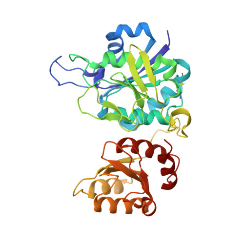
Crystal structure of Escherichia coli L-asparaginase, an enzyme used in cancer therapy.

Swain, A.L.Jaskolski, M.Housset, D.Rao, J.K.Wlodawer, A.

(1993) Proc Natl Acad Sci U S A 90: 1474-1478

  • DOI: https://doi.org/10.1073/pnas.90.4.1474
  • Primary Citation of Related Structures:  
    3ECA

  • PubMed Abstract: 

    The crystal structure of Escherichia coli asparaginase II (EC 3.5.1.1), a drug (Elspar) used for the treatment of acute lymphoblastic leukemia, has been determined at 2.3 A resolution by using data from a single heavy atom derivative in combination with molecular replacement. The atomic model was refined to an R factor of 0.143. This enzyme, active as a homotetramer with 222 symmetry, belongs to the class of alpha/beta proteins. Each subunit has two domains with unique topological features. On the basis of present structural evidence consistent with previous biochemical studies, we propose locations for the active sites between the N- and C-terminal domains belonging to different subunits and postulate a catalytic role for Thr-89.


  • Organizational Affiliation

    Macromolecular Structure Laboratory, National Cancer Institute-Frederick Cancer Research and Development Center, MD 21702-1201.


Macromolecules
Find similar proteins by:  (by identity cutoff)  |  3D Structure
Entity ID: 1
MoleculeChains Sequence LengthOrganismDetailsImage
L-asparaginase 2
A, B, C, D
326Escherichia coliMutation(s): 0 
EC: 3.5.1.1
UniProt
Find proteins for P00805 (Escherichia coli (strain K12))
Explore P00805 
Go to UniProtKB:  P00805
Entity Groups  
Sequence Clusters30% Identity50% Identity70% Identity90% Identity95% Identity100% Identity
UniProt GroupP00805
Sequence Annotations
Expand
  • Reference Sequence
Experimental Data & Validation

Experimental Data

  • Method: X-RAY DIFFRACTION
  • Resolution: 2.40 Å
  • R-Value Work: 0.111 
  • R-Value Observed: 0.111 
  • Space Group: P 1 21 1
Unit Cell:
Length ( Å )Angle ( ˚ )
a = 76.7α = 90
b = 96.1β = 97.1
c = 111.3γ = 90
Software Package:
Software NamePurpose
REFMACrefinement
X-PLORmodel building
X-PLORphasing
d*TREKdata reduction
d*TREKdata scaling

Structure Validation

View Full Validation Report


Entry History 

Deposition Data

Revision History  (Full details and data files)

  • Version 1.0: 1993-10-31
    Type: Initial release
  • Version 1.1: 2008-03-03
    Changes: Version format compliance
  • Version 1.2: 2011-07-13
    Changes: Version format compliance
  • Version 1.3: 2017-11-29
    Changes: Derived calculations, Other
  • Version 2.0: 2020-07-29
    Type: Coordinate replacement
    Reason: Sequence discrepancy
    Changes: Advisory, Atomic model, Data collection, Database references, Derived calculations, Experimental preparation, Other, Polymer sequence, Refinement description, Source and taxonomy, Structure summary