3EC3

Crystal structure of the bb fragment of ERp72


Experimental Data Snapshot

  • Method: X-RAY DIFFRACTION
  • Resolution: 1.92 Å
  • R-Value Free: 0.236 
  • R-Value Work: 0.188 
  • R-Value Observed: 0.190 

wwPDB Validation   3D Report Full Report


This is version 1.2 of the entry. See complete history


Literature

Structure of the Noncatalytic Domains and Global Fold of the Protein Disulfide Isomerase ERp72.

Kozlov, G.Maattanen, P.Schrag, J.D.Hura, G.L.Gabrielli, L.Cygler, M.Thomas, D.Y.Gehring, K.

(2009) Structure 17: 651-659

  • DOI: https://doi.org/10.1016/j.str.2009.02.016
  • Primary Citation of Related Structures:  
    3EC3

  • PubMed Abstract: 

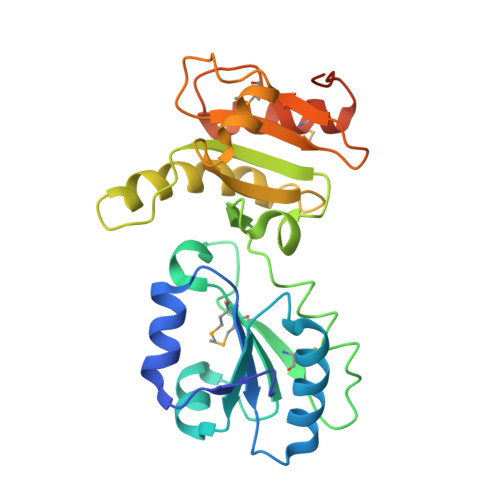
    Protein disulfide isomerases are a family of proteins that catalyze the oxidation and isomerization of disulfide bonds in newly synthesized proteins in the endoplasmic reticulum. The family includes general enzymes such as PDI that recognize unfolded proteins, and others that are selective for specific classes of proteins. Here, we report the X-ray crystal structure of central non-catalytic domains of a specific isomerase, ERp72 (also called CaBP2 and protein disulfide-isomerase A4) from Rattus norvegicus. The structure reveals strong similarity to ERp57, a PDI-family member that interacts with the lectin-like chaperones calnexin and calreticulin but, unexpectedly, ERp72 does not interact with calnexin as shown by isothermal titration calorimetry and nuclear magnetic resonance (NMR) spectroscopy. Small-angle X-ray scattering (SAXS) of ERp72 was used to develop models of the full-length protein using both rigid body refinement and ab initio simulated annealing of dummy atoms. The two methods show excellent agreement and define the relative positions of the five thioredoxin-like domains of ERp72 and potential substrate or chaperone binding sites.


  • Organizational Affiliation

    Department of Biochemistry, McGill University, 3655 Promenade Sir William Osler, Montréal, Québec, Canada.


Macromolecules
Find similar proteins by:  (by identity cutoff)  |  3D Structure
Entity ID: 1
MoleculeChains Sequence LengthOrganismDetailsImage
Protein disulfide-isomerase A4
A, B
250Rattus norvegicusMutation(s): 1 
Gene Names: Pdia4Cabp2Erp70
EC: 5.3.4.1
UniProt
Find proteins for P38659 (Rattus norvegicus)
Explore P38659 
Go to UniProtKB:  P38659
Entity Groups  
Sequence Clusters30% Identity50% Identity70% Identity90% Identity95% Identity100% Identity
UniProt GroupP38659
Sequence Annotations
Expand
  • Reference Sequence
Small Molecules
Modified Residues  1 Unique
IDChains TypeFormula2D DiagramParent
MSE
Query on MSE
A, B
L-PEPTIDE LINKINGC5 H11 N O2 SeMET
Experimental Data & Validation

Experimental Data

  • Method: X-RAY DIFFRACTION
  • Resolution: 1.92 Å
  • R-Value Free: 0.236 
  • R-Value Work: 0.188 
  • R-Value Observed: 0.190 
  • Space Group: P 31
Unit Cell:
Length ( Å )Angle ( ˚ )
a = 62.171α = 90
b = 62.171β = 90
c = 135.004γ = 120
Software Package:
Software NamePurpose
ADSCdata collection
SHARPphasing
REFMACrefinement
HKL-2000data reduction
HKL-2000data scaling

Structure Validation

View Full Validation Report


Entry History 

Deposition Data

Revision History  (Full details and data files)

  • Version 1.0: 2009-04-14
    Type: Initial release
  • Version 1.1: 2011-07-13
    Changes: Advisory, Version format compliance
  • Version 1.2: 2021-10-20
    Changes: Database references, Derived calculations