


Crystal Structure of the Complex of C-lobe of Lactoferrin with Nimesulide at 2.7 A Resolution
Mir, R., Balaji, K., Vikram, G., Sinha, M., Singh, N., Bhushan, A., Kaur, P., Sharma, S., Singh, T.P.To be published.
Experimental Data Snapshot
Entity ID: 1 | |||||
|---|---|---|---|---|---|
| Molecule | Chains | Sequence Length | Organism | Details | Image |
| Lactotransferrin' | 345 | Bos taurus | Mutation(s): 0 EC: 3.4.21 | 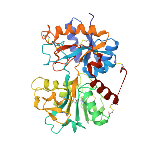 | |
UniProt | |||||
Find proteins for P24627 (Bos taurus) Explore P24627 Go to UniProtKB: P24627 | |||||
Entity Groups | |||||
| Sequence Clusters | 30% Identity50% Identity70% Identity90% Identity95% Identity100% Identity | ||||
| UniProt Group | P24627 | ||||
Sequence AnnotationsExpand | |||||
| |||||
| Ligands 5 Unique | |||||
|---|---|---|---|---|---|
| ID | Chains | Name / Formula / InChI Key | 2D Diagram | 3D Interactions | |
| NIM Query on NIM | E [auth A] | 4-NITRO-2-PHENOXYMETHANESULFONANILIDE C13 H12 N2 O5 S HYWYRSMBCFDLJT-UHFFFAOYSA-N |  | ||
| SO4 Query on SO4 | J [auth A] | SULFATE ION O4 S QAOWNCQODCNURD-UHFFFAOYSA-L |  | ||
| ZN Query on ZN | H [auth A], I [auth A] | ZINC ION Zn PTFCDOFLOPIGGS-UHFFFAOYSA-N |  | ||
| CO3 Query on CO3 | G [auth A] | CARBONATE ION C O3 BVKZGUZCCUSVTD-UHFFFAOYSA-L |  | ||
| FE Query on FE | F [auth A] | FE (III) ION Fe VTLYFUHAOXGGBS-UHFFFAOYSA-N |  | ||
| Length ( Å ) | Angle ( ˚ ) |
|---|---|
| a = 63.57 | α = 90 |
| b = 50.35 | β = 107.9 |
| c = 65.96 | γ = 90 |
| Software Name | Purpose |
|---|---|
| DENZO | data reduction |
| MOLREP | phasing |
| CNS | refinement |
| AUTOMAR | data reduction |
| SCALEPACK | data scaling |
RCSB PDB (citation) is hosted by
RCSB PDB is a member of the


