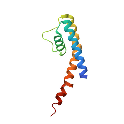
3E8G

Crystal Structure of the the open NaK channel-Na+/Ca2+ complex


Experimental Data Snapshot

  • Method: X-RAY DIFFRACTION
  • Resolution: 2.00 Å
  • R-Value Free: 0.235 
  • R-Value Work: 0.202 

wwPDB Validation   3D Report Full Report


This is version 1.4 of the entry. See complete history


Literature

Structural analysis of ion selectivity in the NaK channel

Alam, A.Jiang, Y.

(2009) Nat Struct Mol Biol 16: 35-41

  • DOI: https://doi.org/10.1038/nsmb.1537
  • Primary Citation of Related Structures:  
    3E83, 3E89, 3E8B, 3E8F, 3E8G, 3E8H

  • PubMed Abstract: 

    Here we present a detailed characterization of ion binding in the NaK pore using the high-resolution structures of NaK in complex with various cations. These structures reveal four ion binding sites with similar chemical environments but vastly different ion preference. The most nonselective of all is site 3, which is formed exclusively by backbone carbonyl oxygen atoms and resides deep within the selectivity filter. Additionally, four water molecules in combination with four backbone carbonyl oxygen atoms are seen to participate in K(+) and Rb(+) ion chelation, at both the external entrance and the vestibule of the NaK filter, confirming the channel's preference for an octahedral ligand configuration for K(+) and Rb(+) binding. In contrast, Na(+) binding in the NaK filter, particularly at site 4, utilizes a pyramidal ligand configuration that requires the participation of a water molecule in the cavity. Therefore, the ability of the NaK filter to bind both Na(+) and K(+) ions seemingly arises from the ions' ability to use the existing environment in unique ways, rather than from any structural rearrangements of the filter itself.


  • Organizational Affiliation

    Department of Physiology, University of Texas Southwestern Medical Center, 5323 Harry Hines Blvd, Dallas, Texas 75390-9040, USA.


Macromolecules
Find similar proteins by:  (by identity cutoff)  |  3D Structure
Entity ID: 1
MoleculeChains Sequence LengthOrganismDetailsImage
Potassium channel protein
A, B
96Bacillus cereusMutation(s): 0 
Membrane Entity: Yes 
UniProt
Find proteins for Q81HW2 (Bacillus cereus (strain ATCC 14579 / DSM 31 / CCUG 7414 / JCM 2152 / NBRC 15305 / NCIMB 9373 / NCTC 2599 / NRRL B-3711))
Explore Q81HW2 
Go to UniProtKB:  Q81HW2
Entity Groups  
Sequence Clusters30% Identity50% Identity70% Identity90% Identity95% Identity100% Identity
UniProt GroupQ81HW2
Sequence Annotations
Expand
  • Reference Sequence
Small Molecules
Ligands 4 Unique
IDChains Name / Formula / InChI Key2D Diagram3D Interactions
CS
Query on CS

Download Ideal Coordinates CCD File 
E [auth A],
J [auth B]
CESIUM ION
Cs
NCMHKCKGHRPLCM-UHFFFAOYSA-N
MPD
Query on MPD

Download Ideal Coordinates CCD File 
C [auth A],
D [auth A],
I [auth B]
(4S)-2-METHYL-2,4-PENTANEDIOL
C6 H14 O2
SVTBMSDMJJWYQN-YFKPBYRVSA-N
CA
Query on CA

Download Ideal Coordinates CCD File 
F [auth A],
K [auth B]
CALCIUM ION
Ca
BHPQYMZQTOCNFJ-UHFFFAOYSA-N
NA
Query on NA

Download Ideal Coordinates CCD File 
G [auth A],
H [auth A],
L [auth B],
M [auth B]
SODIUM ION
Na
FKNQFGJONOIPTF-UHFFFAOYSA-N
Experimental Data & Validation

Experimental Data

  • Method: X-RAY DIFFRACTION
  • Resolution: 2.00 Å
  • R-Value Free: 0.235 
  • R-Value Work: 0.202 
  • Space Group: I 4
Unit Cell:
Length ( Å )Angle ( ˚ )
a = 67.99α = 90
b = 67.99β = 90
c = 89.168γ = 90
Software Package:
Software NamePurpose
DENZOdata reduction
SCALEPACKdata scaling
MOLREPphasing
CNSrefinement
PDB_EXTRACTdata extraction

Structure Validation

View Full Validation Report


Entry History 

Deposition Data

Revision History  (Full details and data files)

  • Version 1.0: 2008-12-23
    Type: Initial release
  • Version 1.1: 2011-07-13
    Changes: Version format compliance
  • Version 1.2: 2011-11-16
    Changes: Atomic model
  • Version 1.3: 2017-10-25
    Changes: Refinement description
  • Version 1.4: 2024-02-21
    Changes: Data collection, Database references, Derived calculations