3E4D

Structural and Kinetic Study of an S-Formylglutathione Hydrolase from Agrobacterium tumefaciens


Experimental Data Snapshot

  • Method: X-RAY DIFFRACTION
  • Resolution: 2.01 Å
  • R-Value Free: 0.219 
  • R-Value Work: 0.181 
  • R-Value Observed: 0.183 

wwPDB Validation   3D Report Full Report


This is version 1.4 of the entry. See complete history


Literature

The structure of a putative S-formylglutathione hydrolase from Agrobacterium tumefaciens

van Straaten, K.E.Gonzalez, C.F.Valladares, R.B.Xu, X.Savchenko, A.V.Sanders, D.A.

(2009) Protein Sci 18: 2196-2202

  • DOI: https://doi.org/10.1002/pro.216
  • Primary Citation of Related Structures:  
    3E4D

  • PubMed Abstract: 

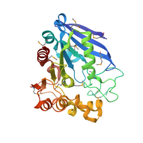
    The structure of the Atu1476 protein from Agrobacterium tumefaciens was determined at 2 A resolution. The crystal structure and biochemical characterization of this enzyme support the conclusion that this protein is an S-formylglutathione hydrolase (AtuSFGH). The three-dimensional structure of AtuSFGH contains the alpha/beta hydrolase fold topology and exists as a homo-dimer. Contacts between the two monomers in the dimer are formed both by hydrogen bonds and salt bridges. Biochemical characterization reveals that AtuSFGH hydrolyzes C--O bonds with high affinity toward short to medium chain esters, unlike the other known SFGHs which have greater affinity toward shorter chained esters. A potential role for Cys54 in regulation of enzyme activity through S-glutathionylation is also proposed.


  • Organizational Affiliation

    Department of Chemistry, University of Saskatchewan, Saskatoon, SK, Canada.


Macromolecules
Find similar proteins by:  (by identity cutoff)  |  3D Structure
Entity ID: 1
MoleculeChains Sequence LengthOrganismDetailsImage
Esterase D278Agrobacterium fabrum str. C58Mutation(s): 0 
Gene Names: AGR_C_2723Atu1476
UniProt
Find proteins for A9CJ11 (Agrobacterium fabrum (strain C58 / ATCC 33970))
Explore A9CJ11 
Go to UniProtKB:  A9CJ11
Entity Groups  
Sequence Clusters30% Identity50% Identity70% Identity90% Identity95% Identity100% Identity
UniProt GroupA9CJ11
Sequence Annotations
Expand
  • Reference Sequence
Small Molecules
Ligands 2 Unique
IDChains Name / Formula / InChI Key2D Diagram3D Interactions
CL
Query on CL

Download Ideal Coordinates CCD File 
H [auth A]
I [auth A]
K [auth B]
L [auth B]
N [auth C]
H [auth A],
I [auth A],
K [auth B],
L [auth B],
N [auth C],
O [auth C],
Q [auth E],
R [auth E],
T [auth D],
U [auth D],
W [auth F],
X [auth F]
CHLORIDE ION
Cl
VEXZGXHMUGYJMC-UHFFFAOYSA-M
MG
Query on MG

Download Ideal Coordinates CCD File 
G [auth A]
J [auth B]
M [auth C]
P [auth E]
S [auth D]
G [auth A],
J [auth B],
M [auth C],
P [auth E],
S [auth D],
V [auth F]
MAGNESIUM ION
Mg
JLVVSXFLKOJNIY-UHFFFAOYSA-N
Modified Residues  1 Unique
IDChains TypeFormula2D DiagramParent
MSE
Query on MSE
A
B
C
D [auth E]
E [auth D]
A,
B,
C,
D [auth E],
E [auth D],
F
L-PEPTIDE LINKINGC5 H11 N O2 SeMET
Experimental Data & Validation

Experimental Data

  • Method: X-RAY DIFFRACTION
  • Resolution: 2.01 Å
  • R-Value Free: 0.219 
  • R-Value Work: 0.181 
  • R-Value Observed: 0.183 
  • Space Group: P 1
Unit Cell:
Length ( Å )Angle ( ˚ )
a = 68.433α = 92.41
b = 68.949β = 99.79
c = 95.648γ = 98.47
Software Package:
Software NamePurpose
DENZOdata reduction
SCALEPACKdata scaling
REFMACrefinement
PDB_EXTRACTdata extraction
HKL-2000data collection
MrBUMPphasing

Structure Validation

View Full Validation Report


Entry History 

Deposition Data

Revision History  (Full details and data files)

  • Version 1.0: 2009-08-18
    Type: Initial release
  • Version 1.1: 2011-07-13
    Changes: Version format compliance
  • Version 1.2: 2017-10-25
    Changes: Advisory, Refinement description
  • Version 1.3: 2023-08-30
    Changes: Advisory, Data collection, Database references, Derived calculations, Refinement description
  • Version 1.4: 2023-11-15
    Changes: Data collection