3E42

Q138F HincII bound to GTCGAC and Ca2+ (cocrystallized)


Experimental Data Snapshot

  • Method: X-RAY DIFFRACTION
  • Resolution: 2.68 Å
  • R-Value Free: 0.283 
  • R-Value Work: 0.188 
  • R-Value Observed: 0.192 

wwPDB Validation   3D Report Full Report


This is version 1.3 of the entry. See complete history


Literature

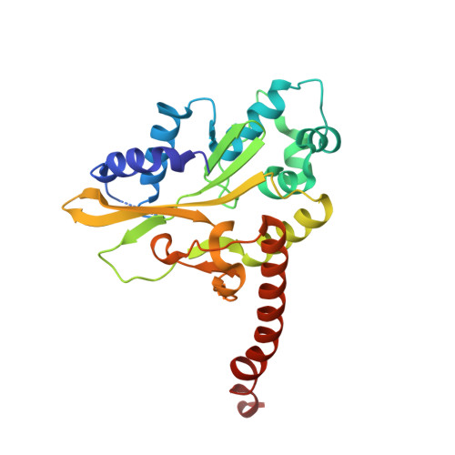
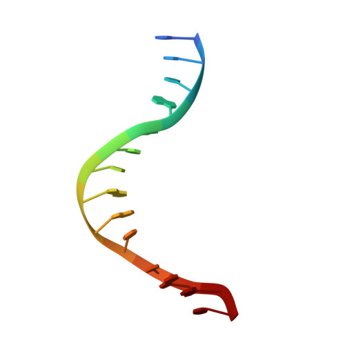
DNA distortion and specificity in a sequence-specific endonuclease.

Babic, A.C.Little, E.J.Manohar, V.M.Bitinaite, J.Horton, N.C.

(2008) J Mol Biol 383: 186-204

  • DOI: https://doi.org/10.1016/j.jmb.2008.08.032
  • Primary Citation of Related Structures:  
    3E3Y, 3E40, 3E41, 3E42, 3E43, 3E44, 3E45

  • PubMed Abstract: 

    Five new structures of the Q138F HincII enzyme bound to a total of three different DNA sequences and three different metal ions (Ca(2+), Mg(2+), and Mn(2+)) are presented. While previous structures were produced from soaking Ca(2+) into preformed Q138F HincII/DNA crystals, the new structures are derived from cocrystallization with Ca(2+), Mg(2+), or Mn(2+). The Mn(2)(+)-bound structure provides the first view of a product complex of Q138F HincII with cleaved DNA. Binding studies and a crystal structure show how Ca(2+) allows trapping of a Q138F HincII complex with noncognate DNA in a catalytically incompetent conformation. Many Q138F HincII/DNA structures show asymmetry, despite the binding of a symmetric substrate by a symmetric enzyme. The various complexes are fit into a model describing the different conformations of the DNA-bound enzyme and show how DNA conformational energetics determine DNA-cleavage rates by the Q138F HincII enzyme.


  • Organizational Affiliation

    Department of Biochemistry and Molecular Biophysics, University of Arizona, Tucson, AZ 85721, USA.


Macromolecules

Find similar proteins by:  (by identity cutoff)  |  3D Structure
Entity ID: 1
MoleculeChains Sequence LengthOrganismDetailsImage
Type-2 restriction enzyme HindII
A, B
257Haemophilus influenzaeMutation(s): 1 
Gene Names: hindIIRHI0512
EC: 3.1.21.4
UniProt
Find proteins for P17743 (Haemophilus influenzae)
Explore P17743 
Go to UniProtKB:  P17743
Entity Groups  
Sequence Clusters30% Identity50% Identity70% Identity90% Identity95% Identity100% Identity
UniProt GroupP17743
Sequence Annotations
Expand
  • Reference Sequence

Find similar nucleic acids by:  Sequence   |   3D Structure  

Entity ID: 2
MoleculeChains LengthOrganismImage
5'-D(*DGP*DCP*DCP*DGP*DGP*DTP*DCP*DGP*DAP*DCP*DCP*DGP*DGP*DC)-3'C [auth E],
D [auth F]
14N/A
Sequence Annotations
Expand
  • Reference Sequence
Experimental Data & Validation

Experimental Data

  • Method: X-RAY DIFFRACTION
  • Resolution: 2.68 Å
  • R-Value Free: 0.283 
  • R-Value Work: 0.188 
  • R-Value Observed: 0.192 
  • Space Group: P 1 21 1
Unit Cell:
Length ( Å )Angle ( ˚ )
a = 50.03α = 90
b = 90.14β = 104.4
c = 66.44γ = 90
Software Package:
Software NamePurpose
PHENIXrefinement
PDB_EXTRACTdata extraction
Blu-Icedata collection
MOSFLMdata reduction
SCALAdata scaling
CNSphasing

Structure Validation

View Full Validation Report


Entry History 

Deposition Data

Revision History  (Full details and data files)

  • Version 1.0: 2008-08-26
    Type: Initial release
  • Version 1.1: 2011-07-13
    Changes: Version format compliance
  • Version 1.2: 2021-10-20
    Changes: Database references, Derived calculations
  • Version 1.3: 2024-02-21
    Changes: Data collection