3DVP

Pak1 peptide bound LC8


Experimental Data Snapshot

  • Method: X-RAY DIFFRACTION
  • Resolution: 2.50 Å
  • R-Value Free: 0.263 
  • R-Value Work: 0.204 
  • R-Value Observed: 0.207 

wwPDB Validation   3D Report Full Report


This is version 1.5 of the entry. See complete history


Literature

Biochemical and structural characterization of the Pak1-LC8 interaction.

Lightcap, C.M.Sun, S.Lear, J.D.Rodeck, U.Polenova, T.Williams, J.C.

(2008) J Biol Chem 283: 27314-27324

  • DOI: https://doi.org/10.1074/jbc.M800758200
  • Primary Citation of Related Structures:  
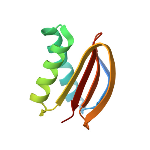
    3DVH, 3DVP, 3DVT

  • PubMed Abstract: 

    Pak1 (p21-activated kinase-1) and the dynein light chain, LC8, are overexpressed in breast cancer, and their direct interaction has been proposed to regulate tumor cell survival. These effects have been attributed in part to Pak1-mediated phosphorylation of LC8 at serine 88. However, LC8 is homodimeric, which renders Ser(88) inaccessible. Moreover, Pak1 does not contain a canonical LC8 binding sequence compared with other characterized LC8 binding sequences. Together, these observations raise the question whether the Pak1/LC8 interaction is distinct (i.e. enabled by a unique interface independent of LC8 dimerization). Herein, we present results from biochemical, NMR, and crystallographic studies that show that Pak1 (residues 212-222) binds to LC8 along the same groove as canonical LC8 interaction partners (e.g. nNOS and BimL). Using LC8 point mutants K36P and T67A, we were able to differentiate Pak1 from canonical LC8 binding sequences and identify a key hydrogen bond network that compensates for the loss of the conserved glutamine in the consensus sequence. We also show that the target binding interface formed through LC8 dimerization is required to bind to Pak1 and precludes phosphorylation of LC8 at Ser(88). Consistent with this observation, in vitro phosphorylation assays using activated Pak1 fail to phosphorylate LC8. Although these results define structural details of the Pak1/LC8 interaction and suggest a hierarchy of target binding affinities, they do not support the current model whereby Pak1 binds to and subsequently phosphorylates LC8 to promote anchorage-independent growth. Rather, they suggest that LC8 binding modulates Pak1 activity and/or nuclear localization.


  • Organizational Affiliation

    Department of Biochemistry and Molecular Biology, Thomas Jefferson University, Philadelphia, Pennsylvania 19107, USA.


Macromolecules
Find similar proteins by:  (by identity cutoff)  |  3D Structure
Entity ID: 1
MoleculeChains Sequence LengthOrganismDetailsImage
Dynein light chain 1, cytoplasmic
A, B
91Drosophila melanogasterMutation(s): 1 
Gene Names: ctpCdlc1ddlc1CG6998
UniProt
Find proteins for Q24117 (Drosophila melanogaster)
Explore Q24117 
Go to UniProtKB:  Q24117
Entity Groups  
Sequence Clusters30% Identity50% Identity70% Identity90% Identity95% Identity100% Identity
UniProt GroupQ24117
Sequence Annotations
Expand
  • Reference Sequence

Find similar proteins by:  Sequence   |   3D Structure  

Entity ID: 2
MoleculeChains Sequence LengthOrganismDetailsImage
P21 activated Kinase peptide
C, D
10N/AMutation(s): 0 
UniProt & NIH Common Fund Data Resources
Find proteins for Q13153 (Homo sapiens)
Explore Q13153 
Go to UniProtKB:  Q13153
PHAROS:  Q13153
GTEx:  ENSG00000149269 
Entity Groups  
Sequence Clusters30% Identity50% Identity70% Identity90% Identity95% Identity100% Identity
UniProt GroupQ13153
Sequence Annotations
Expand
  • Reference Sequence
Experimental Data & Validation

Experimental Data

  • Method: X-RAY DIFFRACTION
  • Resolution: 2.50 Å
  • R-Value Free: 0.263 
  • R-Value Work: 0.204 
  • R-Value Observed: 0.207 
  • Space Group: P 21 21 21
Unit Cell:
Length ( Å )Angle ( ˚ )
a = 47.404α = 90
b = 57.378β = 90
c = 64.801γ = 90
Software Package:
Software NamePurpose
REFMACrefinement
CrysalisProdata collection
CrysalisProdata reduction
SCALAdata scaling
PHASERphasing

Structure Validation

View Full Validation Report


Entry History 

Deposition Data

Revision History  (Full details and data files)

  • Version 1.0: 2009-01-20
    Type: Initial release
  • Version 1.1: 2011-07-13
    Changes: Advisory, Version format compliance
  • Version 1.2: 2017-10-25
    Changes: Refinement description
  • Version 1.3: 2018-04-04
    Changes: Data collection
  • Version 1.4: 2021-10-20
    Changes: Database references
  • Version 1.5: 2024-02-21
    Changes: Data collection