3DK8

Catalytic cycle of human glutathione reductase near 1 A resolution


Experimental Data Snapshot

  • Method: X-RAY DIFFRACTION
  • Resolution: 1.10 Å
  • R-Value Free: 0.157 
  • R-Value Observed: 0.124 

wwPDB Validation   3D Report Full Report


Ligand Structure Quality Assessment 


This is version 1.4 of the entry. See complete history


Literature

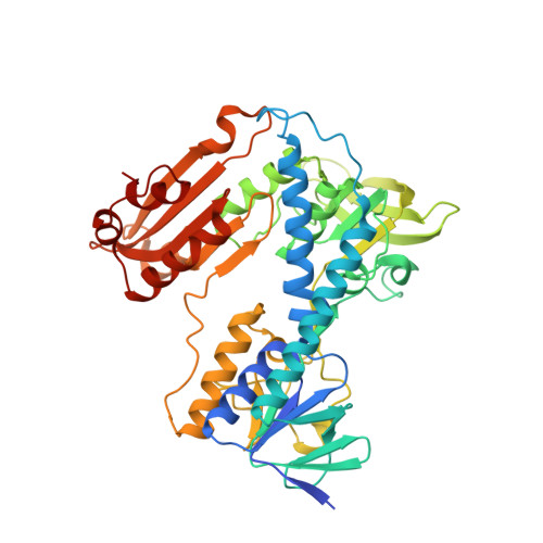
Catalytic cycle of human glutathione reductase near 1 A resolution.

Berkholz, D.S.Faber, H.R.Savvides, S.N.Karplus, P.A.

(2008) J Mol Biol 382: 371-384

  • DOI: https://doi.org/10.1016/j.jmb.2008.06.083
  • Primary Citation of Related Structures:  
    3DJG, 3DJJ, 3DK4, 3DK8, 3DK9

  • PubMed Abstract: 

    Efficient enzyme catalysis depends on exquisite details of structure beyond those resolvable in typical medium- and high-resolution crystallographic analyses. Here we report synchrotron-based cryocrystallographic studies of natural substrate complexes of the flavoenzyme human glutathione reductase (GR) at nominal resolutions between 1.1 and 0.95 A that reveal new aspects of its mechanism. Compression in the active site causes overlapping van der Waals radii and distortion in the nicotinamide ring of the NADPH substrate, which enhances catalysis via stereoelectronic effects. The bound NADPH and redox-active disulfide are positioned optimally on opposite sides of the flavin for a 1,2-addition across a flavin double bond. The new structures extend earlier observations to reveal that the redox-active disulfide loop in GR is an extreme case of sequential peptide bonds systematically deviating from planarity--a net deviation of 53 degrees across five residues. But this apparent strain is not a factor in catalysis, as it is present in both oxidized and reduced structures. Intriguingly, the flavin bond lengths in oxidized GR are intermediate between those expected for oxidized and reduced flavin, but we present evidence that this may not be due to the protein environment but instead due to partial synchrotron reduction of the flavin by the synchrotron beam. Finally, of more general relevance, we present evidence that the structures of synchrotron-reduced disulfide bonds cannot generally be used as reliable models for naturally reduced disulfide bonds.


  • Organizational Affiliation

    Department of Biochemistry and Biophysics, Oregon State University, 2011 ALS, Corvallis, OR 97331-7305, USA.


Macromolecules
Find similar proteins by:  (by identity cutoff)  |  3D Structure
Entity ID: 1
MoleculeChains Sequence LengthOrganismDetailsImage
Glutathione reductase478Homo sapiensMutation(s): 0 
Gene Names: GSRGLURGRD1
EC: 1.8.1.7
UniProt & NIH Common Fund Data Resources
Find proteins for P00390 (Homo sapiens)
Explore P00390 
Go to UniProtKB:  P00390
PHAROS:  P00390
GTEx:  ENSG00000104687 
Entity Groups  
Sequence Clusters30% Identity50% Identity70% Identity90% Identity95% Identity100% Identity
UniProt GroupP00390
Sequence Annotations
Expand
  • Reference Sequence
Small Molecules
Ligands 4 Unique
IDChains Name / Formula / InChI Key2D Diagram3D Interactions
FAD
Query on FAD

Download Ideal Coordinates CCD File 
D [auth A]FLAVIN-ADENINE DINUCLEOTIDE
C27 H33 N9 O15 P2
VWWQXMAJTJZDQX-UYBVJOGSSA-N
GSH
Query on GSH

Download Ideal Coordinates CCD File 
E [auth A],
F [auth A]
GLUTATHIONE
C10 H17 N3 O6 S
RWSXRVCMGQZWBV-WDSKDSINSA-N
SO4
Query on SO4

Download Ideal Coordinates CCD File 
B [auth A],
C [auth A]
SULFATE ION
O4 S
QAOWNCQODCNURD-UHFFFAOYSA-L
GOL
Query on GOL

Download Ideal Coordinates CCD File 
G [auth A],
H [auth A],
I [auth A],
J [auth A]
GLYCEROL
C3 H8 O3
PEDCQBHIVMGVHV-UHFFFAOYSA-N
Experimental Data & Validation

Experimental Data

  • Method: X-RAY DIFFRACTION
  • Resolution: 1.10 Å
  • R-Value Free: 0.157 
  • R-Value Observed: 0.124 
  • Space Group: C 1 2 1
Unit Cell:
Length ( Å )Angle ( ˚ )
a = 120.031α = 90
b = 62.281β = 121.9
c = 83.98γ = 90
Software Package:
Software NamePurpose
DENZOdata reduction
SCALEPACKdata scaling
SHELXrefinement
PDB_EXTRACTdata extraction
CNSphasing
SHELXL-97refinement

Structure Validation

View Full Validation Report


Ligand Structure Quality Assessment 


Entry History 

Deposition Data

Revision History  (Full details and data files)

  • Version 1.0: 2008-08-05
    Type: Initial release
  • Version 1.1: 2011-07-13
    Changes: Non-polymer description, Version format compliance
  • Version 1.2: 2013-12-25
    Changes: Structure summary
  • Version 1.3: 2017-10-25
    Changes: Advisory, Refinement description
  • Version 1.4: 2023-08-30
    Changes: Advisory, Data collection, Database references, Derived calculations, Refinement description